Structural basis for specific RNA recognition by the alternative splicing factor RBM5.
Soni, K., Jagtap, P.K.A., Martinez-Lumbreras, S., Bonnal, S., Geerlof, A., Stehle, R., Simon, B., Valcarcel, J., Sattler, M.(2023) Nat Commun 14: 4233-4233
- PubMed: 37454201 Search on PubMedSearch on PubMed Central
- DOI: https://doi.org/10.1038/s41467-023-39961-w
- Primary Citation Related Structures:
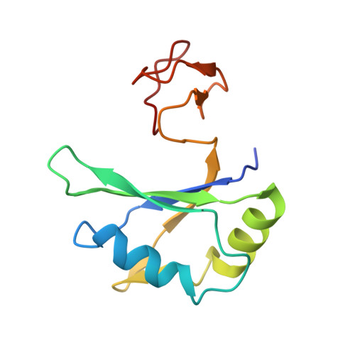
7PCV, 7PDV - PubMed Abstract:
The RNA-binding motif protein RBM5 belongs to a family of multi-domain RNA binding proteins that regulate alternative splicing of genes important for apoptosis and cell proliferation and have been implicated in cancer. RBM5 harbors structural modules for RNA recognition, such as RRM domains and a Zn finger, and protein-protein interactions such as an OCRE domain. Here, we characterize binding of the RBM5 RRM1-ZnF1-RRM2 domains to cis-regulatory RNA elements. A structure of the RRM1-ZnF1 region in complex with RNA shows how the tandem domains cooperate to sandwich target RNA and specifically recognize a GG dinucleotide in a non-canonical fashion. While the RRM1-ZnF1 domains act as a single structural module, RRM2 is connected by a flexible linker and tumbles independently. However, all three domains participate in RNA binding and adopt a closed architecture upon RNA binding. Our data highlight how cooperativity and conformational modularity of multiple RNA binding domains enable the recognition of distinct RNA motifs, thereby contributing to the regulation of alternative splicing. Remarkably, we observe surprising differences in coupling of the RNA binding domains between the closely related homologs RBM5 and RBM10.
- Helmholtz Munich, Molecular Targets and Therapeutics Center, Institute of Structural Biology, Ingolstädter Landstrasse 1, 85764, Neuherberg, Germany.
Organizational Affiliation: