7PAF

Streptococcus pneumoniae choline importer LicB in lipid nanodiscs


Experimental Data Snapshot

  • Method: ELECTRON MICROSCOPY
  • Resolution: 3.75 Å
  • Aggregation State: PARTICLE 
  • Reconstruction Method: SINGLE PARTICLE 

wwPDB Validation   3D Report Full Report


This is version 1.0 of the entry. See complete history


Literature

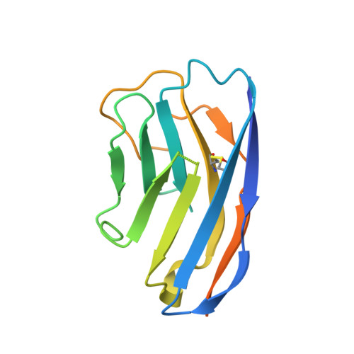
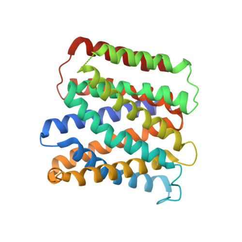
Mechanistic basis of choline import involved in teichoic acids and lipopolysaccharide modification.

Barland, N.Rueff, A.S.Cebrero, G.Hutter, C.A.J.Seeger, M.A.Veening, J.W.Perez, C.

(2022) Sci Adv 8: eabm1122-eabm1122

  • DOI: https://doi.org/10.1126/sciadv.abm1122
  • Primary Citation of Related Structures:  
    7B0K, 7PAF

  • PubMed Abstract: 

    Phosphocholine molecules decorating bacterial cell wall teichoic acids and outer-membrane lipopolysaccharide have fundamental roles in adhesion to host cells, immune evasion, and persistence. Bacteria carrying the operon that performs phosphocholine decoration synthesize phosphocholine after uptake of the choline precursor by LicB, a conserved transporter among divergent species. Streptococcus pneumoniae is a prominent pathogen where phosphocholine decoration plays a fundamental role in virulence. Here, we present cryo-electron microscopy and crystal structures of S. pneumoniae LicB, revealing distinct conformational states and describing architectural and mechanistic elements essential to choline import. Together with in vitro and in vivo functional characterization, we found that LicB displays proton-coupled import activity and promiscuous selectivity involved in adaptation to choline deprivation conditions, and describe LicB inhibition by synthetic nanobodies (sybodies). Our results provide previously unknown insights into the molecular mechanism of a key transporter involved in bacterial pathogenesis and establish a basis for inhibition of the phosphocholine modification pathway across bacterial phyla.


  • Organizational Affiliation

    Biozentrum, University of Basel, Basel 4056, Switzerland.


Macromolecules
Find similar proteins by:  (by identity cutoff)  |  3D Structure
Entity ID: 1
MoleculeChains Sequence LengthOrganismDetailsImage
NanobodyA [auth B],
C
154synthetic constructMutation(s): 0 
Entity Groups  
Sequence Clusters30% Identity50% Identity70% Identity90% Identity95% Identity100% Identity
Sequence Annotations
Expand
  • Reference Sequence
Find similar proteins by:  (by identity cutoff)  |  3D Structure
Entity ID: 2
MoleculeChains Sequence LengthOrganismDetailsImage
LicB proteinB [auth A],
D
292Streptococcus pneumoniae TIGR4Mutation(s): 0 
Gene Names: licBSP_1268
Membrane Entity: Yes 
UniProt
Find proteins for A0A0H2UQH5 (Streptococcus pneumoniae serotype 4 (strain ATCC BAA-334 / TIGR4))
Explore A0A0H2UQH5 
Go to UniProtKB:  A0A0H2UQH5
Entity Groups  
Sequence Clusters30% Identity50% Identity70% Identity90% Identity95% Identity100% Identity
UniProt GroupA0A0H2UQH5
Sequence Annotations
Expand
  • Reference Sequence
Small Molecules
Ligands 1 Unique
IDChains Name / Formula / InChI Key2D Diagram3D Interactions
PGT
Query on PGT

Download Ideal Coordinates CCD File 
E [auth D](1S)-2-{[{[(2R)-2,3-DIHYDROXYPROPYL]OXY}(HYDROXY)PHOSPHORYL]OXY}-1-[(PALMITOYLOXY)METHYL]ETHYL STEARATE
C40 H79 O10 P
KBPVYRBBONZJHF-AMAPPZPBSA-N
Experimental Data & Validation

Experimental Data

  • Method: ELECTRON MICROSCOPY
  • Resolution: 3.75 Å
  • Aggregation State: PARTICLE 
  • Reconstruction Method: SINGLE PARTICLE 
EM Software:
TaskSoftware PackageVersion
RECONSTRUCTIONcryoSPARCv3.2.0

Structure Validation

View Full Validation Report


Entry History & Funding Information

Deposition Data


Funding OrganizationLocationGrant Number
Swiss National Science FoundationSwitzerlandPP00P3_170607

Revision History  (Full details and data files)

  • Version 1.0: 2022-03-16
    Type: Initial release