


Crystal structure of Mycobacterium hassiacum glucosyl-3-phosphoglycerate synthase at pH 5.5 - apo form
Silva, A., Nunes-Costa, D., Empadinhas, N., Barbosa Pereira, P.J., Macedo-Ribeiro, S.To be published.
Experimental Data Snapshot
Starting Model: experimental
View more details
Entity ID: 1 | |||||
|---|---|---|---|---|---|
| Molecule | Chains | Sequence Length | Organism | Details | Image |
| Glucosyl-3-phosphoglycerate synthase | 327 | Mycolicibacterium hassiacum DSM 44199 | Mutation(s): 0 Gene Names: gpgS, C731_3243, MHAS_02845 EC: 2.4.1.266 | 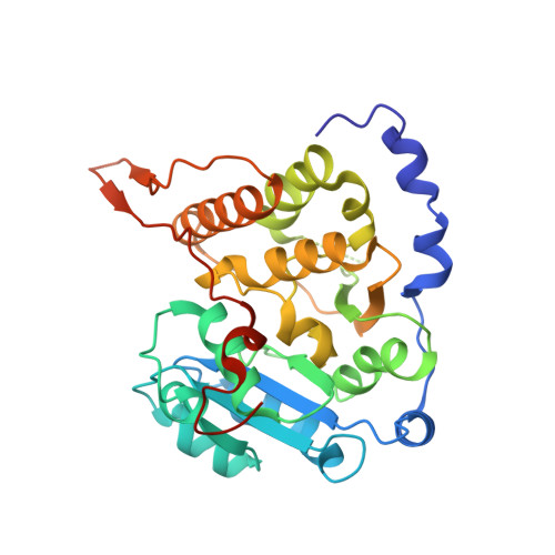 | |
UniProt | |||||
Entity Groups | |||||
| Sequence Clusters | 30% Identity50% Identity70% Identity90% Identity95% Identity100% Identity | ||||
| UniProt Group | K5B7Z4 | ||||
Sequence AnnotationsExpand | |||||
Reference Sequence | |||||
| Ligands 3 Unique | |||||
|---|---|---|---|---|---|
| ID | Chains | Name / Formula / InChI Key | 2D Diagram | 3D Interactions | |
| BGC Download:Ideal Coordinates CCD File | C [auth A] | beta-D-glucopyranose C6 H12 O6 WQZGKKKJIJFFOK-VFUOTHLCSA-N |  | ||
| MLI Download:Ideal Coordinates CCD File | G [auth A] | MALONATE ION C3 H2 O4 OFOBLEOULBTSOW-UHFFFAOYSA-L |  | ||
| CL Download:Ideal Coordinates CCD File | B [auth A], D [auth A], E [auth A], F [auth A] | CHLORIDE ION Cl VEXZGXHMUGYJMC-UHFFFAOYSA-M |  | ||
| Length ( Å ) | Angle ( ˚ ) |
|---|---|
| a = 101.423 | α = 90 |
| b = 101.423 | β = 90 |
| c = 122.625 | γ = 90 |
| Software Name | Purpose |
|---|---|
| PHENIX | refinement |
| XDS | data reduction |
| SCALA | data scaling |
| PHASER | phasing |
| Funding Organization | Location | Grant Number |
|---|---|---|
| Fundacao para a Ciencia e a Tecnologia | Portugal | -- |


