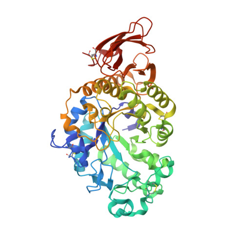
7P4W

Crystal structure of alpha-amylase from Aspergillus oryzae in space group I222


Experimental Data Snapshot

  • Method: X-RAY DIFFRACTION
  • Resolution: 2.28 Å
  • R-Value Free: 0.285 
  • R-Value Work: 0.228 
  • R-Value Observed: 0.231 

wwPDB Validation   3D Report Full Report


This is version 1.2 of the entry. See complete history


Literature

The FUSION protein crystallization screen.

Gorrec, F.Bellini, D.

(2022) J Appl Crystallogr 55: 310-319

  • DOI: https://doi.org/10.1107/S1600576722001765
  • Primary Citation of Related Structures:  
    7P4W, 7P4Z

  • PubMed Abstract: 

    The success and speed of atomic structure determination of biological macromolecules by X-ray crystallography depends critically on the availability of diffraction-quality crystals. However, the process of screening crystallization conditions often consumes large amounts of sample and time. An innovative protein crystallization screen formulation called FUSION has been developed to help with the production of useful crystals. The concept behind the formulation of FUSION was to combine the most efficient components from the three MORPHEUS screens into a single screen using a systematic approach. The resulting formulation integrates 96 unique combinations of crystallization additives. Most of these additives are small molecules and ions frequently found in crystal structures of the Protein Data Bank (PDB), where they bind proteins and complexes. The efficiency of FUSION is demonstrated by obtaining high yields of diffraction-quality crystals for seven different test proteins. In the process, two crystal forms not currently in the PDB for the proteins α-amylase and avidin were discovered.


  • Organizational Affiliation

    Structural Studies, MRC Laboratory of Molecular Biology, Francis Crick Avenue, Cambridge, Cambridgeshire CB2 0QH, United Kingdom.


Macromolecules
Find similar proteins by:  (by identity cutoff)  |  3D Structure
Entity ID: 1
MoleculeChains Sequence LengthOrganismDetailsImage
Alpha-amylase476Aspergillus oryzaeMutation(s): 0 
EC: 3.2.1.1
UniProt
Find proteins for P0C1B3 (Aspergillus oryzae (strain ATCC 42149 / RIB 40))
Explore P0C1B3 
Go to UniProtKB:  P0C1B3
Entity Groups  
Sequence Clusters30% Identity50% Identity70% Identity90% Identity95% Identity100% Identity
UniProt GroupP0C1B3
Sequence Annotations
Expand
  • Reference Sequence
Oligosaccharides

Help

Entity ID: 2
MoleculeChains Length2D Diagram Glycosylation3D Interactions
2-acetamido-2-deoxy-beta-D-glucopyranose-(1-4)-2-acetamido-2-deoxy-beta-D-glucopyranoseB [auth E]2N-Glycosylation
Glycosylation Resources
GlyTouCan:  G42666HT
GlyCosmos:  G42666HT
GlyGen:  G42666HT
Small Molecules
Ligands 2 Unique
IDChains Name / Formula / InChI Key2D Diagram3D Interactions
MES
Query on MES

Download Ideal Coordinates CCD File 
D [auth A]2-(N-MORPHOLINO)-ETHANESULFONIC ACID
C6 H13 N O4 S
SXGZJKUKBWWHRA-UHFFFAOYSA-N
CA
Query on CA

Download Ideal Coordinates CCD File 
C [auth A]CALCIUM ION
Ca
BHPQYMZQTOCNFJ-UHFFFAOYSA-N
Experimental Data & Validation

Experimental Data

  • Method: X-RAY DIFFRACTION
  • Resolution: 2.28 Å
  • R-Value Free: 0.285 
  • R-Value Work: 0.228 
  • R-Value Observed: 0.231 
  • Space Group: I 2 2 2
Unit Cell:
Length ( Å )Angle ( ˚ )
a = 65.725α = 90
b = 140.402β = 90
c = 154.218γ = 90
Software Package:
Software NamePurpose
REFMACrefinement
PHENIXrefinement
DIALSdata reduction
DIALSdata scaling
MOLREPphasing

Structure Validation

View Full Validation Report


Entry History 

Deposition Data

Revision History  (Full details and data files)

  • Version 1.0: 2021-08-25
    Type: Initial release
  • Version 1.1: 2022-05-11
    Changes: Database references
  • Version 1.2: 2024-01-31
    Changes: Data collection, Refinement description