7OZ8

Sulfated host glycan recognition by carbohydrate sulfatases of the human gut microbiota (BT1918_S1_46)


Experimental Data Snapshot

  • Method: X-RAY DIFFRACTION
  • Resolution: 1.95 Å
  • R-Value Free: 0.204 
  • R-Value Work: 0.188 

wwPDB Validation   3D Report Full Report


Ligand Structure Quality Assessment 


This is version 1.2 of the entry. See complete history


Literature

Sulfated glycan recognition by carbohydrate sulfatases of the human gut microbiota.

Luis, A.S.Basle, A.Byrne, D.P.Wright, G.S.A.London, J.A.Jin, C.Karlsson, N.G.Hansson, G.C.Eyers, P.A.Czjzek, M.Barbeyron, T.Yates, E.A.Martens, E.C.Cartmell, A.

(2022) Nat Chem Biol 18: 841-849

  • DOI: https://doi.org/10.1038/s41589-022-01039-x
  • Primary Citation of Related Structures:  
    7OZ8, 7OZ9, 7OZA, 7OZC, 7OZE, 7P24, 7P26

  • PubMed Abstract: 

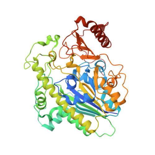
    Sulfated glycans are ubiquitous nutrient sources for microbial communities that have coevolved with eukaryotic hosts. Bacteria metabolize sulfated glycans by deploying carbohydrate sulfatases that remove sulfate esters. Despite the biological importance of sulfatases, the mechanisms underlying their ability to recognize their glycan substrate remain poorly understood. Here, we use structural biology to determine how sulfatases from the human gut microbiota recognize sulfated glycans. We reveal seven new carbohydrate sulfatase structures spanning four S1 sulfatase subfamilies. Structures of S1_16 and S1_46 represent novel structures of these subfamilies. Structures of S1_11 and S1_15 demonstrate how non-conserved regions of the protein drive specificity toward related but distinct glycan targets. Collectively, these data reveal that carbohydrate sulfatases are highly selective for the glycan component of their substrate. These data provide new approaches for probing sulfated glycan metabolism while revealing the roles carbohydrate sulfatases play in host glycan catabolism.


  • Organizational Affiliation

    Department of Microbiology and Immunology, University of Michigan, Ann Arbor, MI, USA. ana.luis@medkem.gu.se.


Macromolecules
Find similar proteins by:  (by identity cutoff)  |  3D Structure
Entity ID: 1
MoleculeChains Sequence LengthOrganismDetailsImage
Choline-sulfataseA [auth AAA]502Bacteroides thetaiotaomicron VPI-5482Mutation(s): 0 
Gene Names: BT_1918
UniProt
Find proteins for Q8A6G6 (Bacteroides thetaiotaomicron (strain ATCC 29148 / DSM 2079 / JCM 5827 / CCUG 10774 / NCTC 10582 / VPI-5482 / E50))
Explore Q8A6G6 
Go to UniProtKB:  Q8A6G6
Entity Groups  
Sequence Clusters30% Identity50% Identity70% Identity90% Identity95% Identity100% Identity
UniProt GroupQ8A6G6
Sequence Annotations
Expand
  • Reference Sequence
Small Molecules
Ligands 2 Unique
IDChains Name / Formula / InChI Key2D Diagram3D Interactions
NGS (Subject of Investigation/LOI)
Query on NGS

Download Ideal Coordinates CCD File 
B [auth AAA]2-acetamido-2-deoxy-6-O-sulfo-beta-D-glucopyranose
C8 H15 N O9 S
WJFVEEAIYIOATH-FMDGEEDCSA-N
ACT
Query on ACT

Download Ideal Coordinates CCD File 
C [auth AAA]ACETATE ION
C2 H3 O2
QTBSBXVTEAMEQO-UHFFFAOYSA-M
Experimental Data & Validation

Experimental Data

  • Method: X-RAY DIFFRACTION
  • Resolution: 1.95 Å
  • R-Value Free: 0.204 
  • R-Value Work: 0.188 
  • Space Group: P 32 2 1
Unit Cell:
Length ( Å )Angle ( ˚ )
a = 56.389α = 90
b = 56.389β = 90
c = 279.763γ = 120
Software Package:
Software NamePurpose
REFMACrefinement
xia2data reduction
Aimlessdata scaling
PHASERphasing

Structure Validation

View Full Validation Report


Ligand Structure Quality Assessment 


Entry History & Funding Information

Deposition Data

  • Released Date: 2022-06-29 
  • Deposition Author(s): Cartmell, A.

Funding OrganizationLocationGrant Number
Wellcome TrustUnited Kingdom--

Revision History  (Full details and data files)

  • Version 1.0: 2022-06-29
    Type: Initial release
  • Version 1.1: 2022-08-10
    Changes: Database references
  • Version 1.2: 2024-01-31
    Changes: Data collection, Refinement description