7OY6 | pdb_00007oy6

Crystal structure of human DYRK1A in complex with ARN25068


Experimental Data Snapshot

  • Method: X-RAY DIFFRACTION
  • Resolution: 2.38 Å
  • R-Value Free: 
    0.270 (Depositor), 0.265 (DCC) 
  • R-Value Work: 
    0.198 (Depositor), 0.208 (DCC) 
  • R-Value Observed: 
    0.202 (Depositor) 

Starting Model: experimental
View more details

wwPDB Validation 3D Report Full Report

Validation slider image for 7OY6

Ligand Structure Quality Assessment 


This is version 1.2 of the entry. See complete history

Literature

ARN25068, a versatile starting point towards triple GSK-3 beta /FYN/DYRK1A inhibitors to tackle tau-related neurological disorders.

Demuro, S.Sauvey, C.Tripathi, S.K.Di Martino, R.M.C.Shi, D.Ortega, J.A.Russo, D.Balboni, B.Giabbai, B.Storici, P.Girotto, S.Abagyan, R.Cavalli, A.

(2022) Eur J Med Chem 229: 114054-114054

  • DOI: https://doi.org/10.1016/j.ejmech.2021.114054
  • Primary Citation Related Structures: 
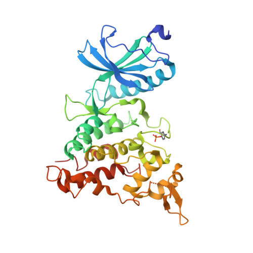
    7OY5, 7OY6

  • PubMed Abstract: 

    The human kinome plays a crucial role in several pathways. Its dysregulation has been linked to diverse central nervous system (CNS)-related disorders with a drastic impact on the aging population. Among them, tauopathies, such as Alzheimer's Disease (AD) and Frontotemporal Lobar degeneration (FTLD-tau), are neurodegenerative disorders pathologically defined by the presence of hyperphosphorylated tau-positive intracellular inclusions known as neurofibrillary tangles (NFTs). Compelling evidence has reported the great potential of the simultaneous modulation of multiple protein kinases (PKs) involved in abnormal tau phosphorylation through a concerted pharmacological approach to achieve a superior therapeutic effect relative to classic "one target, one drug" approaches. Here, we report on the identification and characterization of ARN25068 (4), a low nanomolar and well-balanced dual GSK-3β and FYN inhibitor, which also shows inhibitory activity against DYRK1A, an emerging target in AD and tauopathies. Computational and X-Ray studies highlight compound 4's typical H-bonding pattern of ATP-competitive inhibitors at the binding sites of all three PKs. In a tau phosphorylation assay on Tau0N4R-TM-tGFP U2OS cell line, 4 reduces the extent of tau phosphorylation, promoting tau-stabilized microtubule bundles. In conclusion, this compound emerges as a promising prototype for further SAR explorations to develop potent and well-balanced triple GSK-3β/FYN/DYRK1A inhibitors to tackle tau hyperphosphorylation.


  • Organizational Affiliation
    • Computational and Chemical Biology, Istituto Italiano di Tecnologia, Via Morego 30, 16163, Genoa, Italy; Department of Pharmacy and Biotechnology, University of Bologna, Via Belmeloro 6, 40126, Bologna, Italy.

Macromolecule Content 

  • Total Structure Weight: 42.23 kDa 
  • Atom Count: 2,798 
  • Modeled Residue Count: 343 
  • Deposited Residue Count: 359 
  • Unique protein chains: 1

Macromolecules

Find similar proteins by:|  3D Structure
Entity ID: 1
MoleculeChains  Sequence LengthOrganismDetailsImage
Dual specificity tyrosine-phosphorylation-regulated kinase 1AA [auth C]359Homo sapiensMutation(s): 0 
Gene Names: DYRK1ADYRKMNBMNBH
EC: 2.7.12.1 (PDB Primary Data), 2.7.11.23 (UniProt)
UniProt & NIH Common Fund Data Resources
Find proteins for Q13627 (Homo sapiens)
Explore Q13627 
Go to UniProtKB:  Q13627
PHAROS:  Q13627
GTEx:  ENSG00000157540 
Entity Groups
Sequence Clusters30% Identity50% Identity70% Identity90% Identity95% Identity100% Identity
UniProt GroupQ13627
Sequence Annotations
Expand
Reference Sequence

Small Molecules

Ligands 1 Unique
IDChains Name / Formula / InChI Key2D Diagram3D Interactions
39I
(Subject of Investigation/LOI)

Query on 39I


Download:Ideal Coordinates CCD File
B [auth C]~{N}4-(3-cyclopropyl-1~{H}-pyrazol-5-yl)-~{N}2-(phenylmethyl)thieno[3,2-d]pyrimidine-2,4-diamine
C19 H18 N6 S
TVUWQDLBEOZVOU-UHFFFAOYSA-N
Modified Residues  1 Unique
IDChains TypeFormula2D DiagramParent
PTR
Query on PTR
A [auth C]L-PEPTIDE LINKINGC9 H12 N O6 PTYR

Experimental Data & Validation

Experimental Data

  • Method: X-RAY DIFFRACTION
  • Resolution: 2.38 Å
  • R-Value Free:  0.270 (Depositor), 0.265 (DCC) 
  • R-Value Work:  0.198 (Depositor), 0.208 (DCC) 
  • R-Value Observed: 0.202 (Depositor) 
Space Group: P 42
Unit Cell:
Length ( Å )Angle ( ˚ )
a = 93.65α = 90
b = 93.65β = 90
c = 48.96γ = 90
Software Package:
Software NamePurpose
REFMACrefinement
PDB_EXTRACTdata extraction
iMOSFLMdata reduction
Aimlessdata scaling
PHASERphasing

Structure Validation

View Full Validation Report


Ligand Structure Quality Assessment 


Entry History 

Deposition Data

Revision History  (Full details and data files)

  • Version 1.0: 2022-03-02
    Type: Initial release
  • Version 1.1: 2024-01-31
    Changes: Data collection, Refinement description
  • Version 1.2: 2024-10-23
    Changes: Structure summary