


Structural requirements for the specific binding of CRABP2 to cyclin D3
Pastok, M.W., Tomlinson, C.W.E., Tatum, N.J., Basle, A., Noble, M.E.M., Pohl, E., Endicott, J.A.To be published.
Experimental Data Snapshot
Starting Model: experimental
View more details
wwPDB Validation 3D Report Full Report

Entity ID: 1 | |||||
|---|---|---|---|---|---|
| Molecule | Chains | Sequence Length | Organism | Details | Image |
| Cellular retinoic acid-binding protein 2 | 143 | Homo sapiens | Mutation(s): 2 Gene Names: CRABP2 | 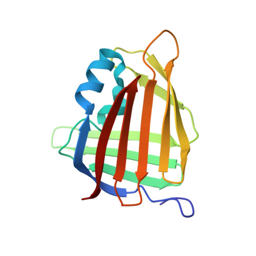 | |
UniProt & NIH Common Fund Data Resources | |||||
PHAROS: P29373 GTEx: ENSG00000143320 | |||||
Entity Groups | |||||
| Sequence Clusters | 30% Identity50% Identity70% Identity90% Identity95% Identity100% Identity | ||||
| UniProt Group | P29373 | ||||
Sequence AnnotationsExpand | |||||
Reference Sequence | |||||
| Ligands 2 Unique | |||||
|---|---|---|---|---|---|
| ID | Chains | Name / Formula / InChI Key | 2D Diagram | 3D Interactions | |
| SO4 Download:Ideal Coordinates CCD File | C [auth A], D [auth A], E [auth A], F [auth A] | SULFATE ION O4 S QAOWNCQODCNURD-UHFFFAOYSA-L |  | ||
| ACT Download:Ideal Coordinates CCD File | B [auth A] | ACETATE ION C2 H3 O2 QTBSBXVTEAMEQO-UHFFFAOYSA-M |  | ||
| Length ( Å ) | Angle ( ˚ ) |
|---|---|
| a = 45.3 | α = 90 |
| b = 92.29 | β = 90 |
| c = 98.76 | γ = 90 |
| Software Name | Purpose |
|---|---|
| XDS | data reduction |
| xia2 | data reduction |
| Aimless | data scaling |
| MolProbity | model building |
| Coot | model building |
| PHASER | phasing |
| REFMAC | refinement |
| Funding Organization | Location | Grant Number |
|---|---|---|
| Medical Research Council (MRC, United Kingdom) | United Kingdom | MR/N009738/1 |


