7OXR

Cryo-EM structure of yeast Sei1 with locking helix deletion


Experimental Data Snapshot

  • Method: ELECTRON MICROSCOPY
  • Resolution: 3.30 Å
  • Aggregation State: PARTICLE 
  • Reconstruction Method: SINGLE PARTICLE 

wwPDB Validation   3D Report Full Report


This is version 1.1 of the entry. See complete history


Literature

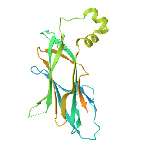
Mechanism of lipid droplet formation by the yeast Sei1/Ldb16 Seipin complex.

Klug, Y.A.Deme, J.C.Corey, R.A.Renne, M.F.Stansfeld, P.J.Lea, S.M.Carvalho, P.

(2021) Nat Commun 12: 5892-5892

  • DOI: https://doi.org/10.1038/s41467-021-26162-6
  • Primary Citation of Related Structures:  
    7OXP, 7OXR

  • PubMed Abstract: 

    Lipid droplets (LDs) are universal lipid storage organelles with a core of neutral lipids, such as triacylglycerols, surrounded by a phospholipid monolayer. This unique architecture is generated during LD biogenesis at endoplasmic reticulum (ER) sites marked by Seipin, a conserved membrane protein mutated in lipodystrophy. Here structural, biochemical and molecular dynamics simulation approaches reveal the mechanism of LD formation by the yeast Seipin Sei1 and its membrane partner Ldb16. We show that Sei1 luminal domain assembles a homooligomeric ring, which, in contrast to other Seipins, is unable to concentrate triacylglycerol. Instead, Sei1 positions Ldb16, which concentrates triacylglycerol within the Sei1 ring through critical hydroxyl residues. Triacylglycerol recruitment to the complex is further promoted by Sei1 transmembrane segments, which also control Ldb16 stability. Thus, we propose that LD assembly by the Sei1/Ldb16 complex, and likely other Seipins, requires sequential triacylglycerol-concentrating steps via distinct elements in the ER membrane and lumen.


  • Organizational Affiliation

    Sir William Dunn School of Pathology, University of Oxford, Oxford, UK.


Macromolecules
Find similar proteins by:  (by identity cutoff)  |  3D Structure
Entity ID: 1
MoleculeChains Sequence LengthOrganismDetailsImage
BJ4_G0032880.mRNA.1.CDS.1
A, B, C, D, E
A, B, C, D, E, F, G, H, I, J
315Saccharomyces cerevisiaeMutation(s): 0 
Gene Names: PACBIOSEQ_LOCUS4180PACBIOSEQ_LOCUS4257PACBIOSEQ_LOCUS4334SCNYR20_0004039900SCP684_0004039500
UniProt
Find proteins for A0A6A5PUS7 (Saccharomyces cerevisiae)
Explore A0A6A5PUS7 
Go to UniProtKB:  A0A6A5PUS7
Entity Groups  
Sequence Clusters30% Identity50% Identity70% Identity90% Identity95% Identity100% Identity
UniProt GroupA0A6A5PUS7
Sequence Annotations
Expand
  • Reference Sequence
Experimental Data & Validation

Experimental Data

  • Method: ELECTRON MICROSCOPY
  • Resolution: 3.30 Å
  • Aggregation State: PARTICLE 
  • Reconstruction Method: SINGLE PARTICLE 

Structure Validation

View Full Validation Report


Entry History & Funding Information

Deposition Data


Funding OrganizationLocationGrant Number
Wellcome TrustUnited Kingdom208361/Z/17/Z
Wellcome TrustUnited Kingdom219477/Z/19/Z
Biotechnology and Biological Sciences Research Council (BBSRC)United KingdomBB/P01948X/1
Biotechnology and Biological Sciences Research Council (BBSRC)United KingdomBB/R002517/1
Biotechnology and Biological Sciences Research Council (BBSRC)United KingdomBB/S003339/1
Medical Research Council (MRC, United Kingdom)United KingdomMR/S009213/1
Biotechnology and Biological Sciences Research Council (BBSRC)United KingdomBB/R018375/1
Wellcome TrustUnited Kingdom202642/Z/16/Z

Revision History  (Full details and data files)

  • Version 1.0: 2021-10-13
    Type: Initial release
  • Version 1.1: 2021-10-20
    Changes: Data collection, Database references