7OVW

Binding domain of botulinum neurotoxin E in complex with GD1a


Experimental Data Snapshot

  • Method: X-RAY DIFFRACTION
  • Resolution: 2.20 Å
  • R-Value Free: 0.271 
  • R-Value Work: 0.216 

wwPDB Validation   3D Report Full Report


This is version 2.1 of the entry. See complete history


Literature

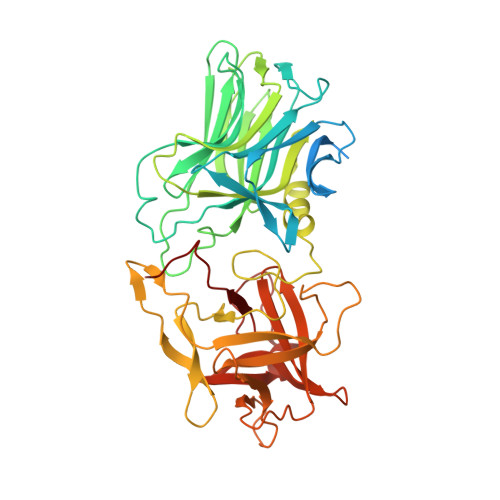
Mechanism of Ganglioside Receptor Recognition by Botulinum Neurotoxin Serotype E.

Masuyer, G.Davies, J.R.Stenmark, P.

(2021) Int J Mol Sci 22

  • DOI: https://doi.org/10.3390/ijms22158315
  • Primary Citation of Related Structures:  
    7OVW

  • PubMed Abstract: 

    The botulinum neurotoxins are potent molecules that are not only responsible for the lethal paralytic disease botulism, but have also been harnessed for therapeutic uses in the treatment of an increasing number of chronic neurological and neuromuscular disorders, in addition to cosmetic applications. The toxins act at the cholinergic nerve terminals thanks to an efficient and specific mechanism of cell recognition which is based on a dual receptor system that involves gangliosides and protein receptors. Binding to surface-anchored gangliosides is the first essential step in this process. Here, we determined the X-ray crystal structure of the binding domain of BoNT/E, a toxin of clinical interest, in complex with its GD1a oligosaccharide receptor. Beyond confirmation of the conserved ganglioside binding site, we identified key interacting residues that are unique to BoNT/E and a significant rearrangement of loop 1228-1237 upon carbohydrate binding. These observations were also supported by thermodynamic measurements of the binding reaction and assessment of ganglioside selectivity by immobilised-receptor binding assays. These results provide a structural basis to understand the specificity of BoNT/E for complex gangliosides.


  • Organizational Affiliation

    Department of Biochemistry and Biophysics, Stockholm University, 10691 Stockholm, Sweden.


Macromolecules
Find similar proteins by:  (by identity cutoff)  |  3D Structure
Entity ID: 1
MoleculeChains Sequence LengthOrganismDetailsImage
Neurotoxin type EA [auth AAA],
B [auth BBB]
468Clostridium botulinumMutation(s): 0 
UniProt
Find proteins for A5H0J8 (Clostridium botulinum)
Explore A5H0J8 
Go to UniProtKB:  A5H0J8
Entity Groups  
Sequence Clusters30% Identity50% Identity70% Identity90% Identity95% Identity100% Identity
UniProt GroupA5H0J8
Sequence Annotations
Expand
  • Reference Sequence
Oligosaccharides

Help

Entity ID: 2
MoleculeChains Length2D Diagram Glycosylation3D Interactions
N-acetyl-alpha-neuraminic acid-(2-3)-beta-D-galactopyranose-(1-3)-2-acetamido-2-deoxy-beta-D-galactopyranose-(1-4)-beta-D-galactopyranose-(1-4)-beta-D-glucopyranoseC [auth GGG]5N/A
Glycosylation Resources
GlyTouCan:  G03277YI
GlyCosmos:  G03277YI
GlyGen:  G03277YI
Entity ID: 3
MoleculeChains Length2D Diagram Glycosylation3D Interactions
N-acetyl-alpha-neuraminic acid-(2-3)-beta-D-galactopyranose-(1-3)-2-acetamido-2-deoxy-beta-D-galactopyranose-(1-4)-beta-D-galactopyranoseD [auth HHH]4N/A
Glycosylation Resources
GlyTouCan:  G02162KR
GlyCosmos:  G02162KR
GlyGen:  G02162KR
Experimental Data & Validation

Experimental Data

  • Method: X-RAY DIFFRACTION
  • Resolution: 2.20 Å
  • R-Value Free: 0.271 
  • R-Value Work: 0.216 
  • Space Group: P 1 21 1
Unit Cell:
Length ( Å )Angle ( ˚ )
a = 69.372α = 90
b = 84.92β = 91.565
c = 79.903γ = 90
Software Package:
Software NamePurpose
REFMACrefinement
XDSdata reduction
Aimlessdata scaling
PHASERphasing

Structure Validation

View Full Validation Report


Entry History & Funding Information

Deposition Data


Funding OrganizationLocationGrant Number
Swedish Research CouncilSweden2018-03406

Revision History  (Full details and data files)

  • Version 1.0: 2021-08-11
    Type: Initial release
  • Version 2.0: 2021-09-29
    Changes: Atomic model, Data collection, Database references
  • Version 2.1: 2024-01-31
    Changes: Data collection, Derived calculations, Refinement description