7OUA

Crystal structure of dimeric chlorite dismutase variant R127K (CCld R127K) from Cyanothece sp. PCC7425


Experimental Data Snapshot

  • Method: X-RAY DIFFRACTION
  • Resolution: 2.09 Å
  • R-Value Free: 0.270 
  • R-Value Work: 0.222 
  • R-Value Observed: 0.225 

wwPDB Validation   3D Report Full Report


This is version 1.2 of the entry. See complete history


Literature

Impact of the dynamics of the catalytic arginine on nitrite and chlorite binding by dimeric chlorite dismutase.

Serra, I.Schmidt, D.Pfanzagl, V.Mlynek, G.Hofbauer, S.Djinovic-Carugo, K.Furtmuller, P.G.Garcia-Rubio, I.Van Doorslaer, S.Obinger, C.

(2021) J Inorg Biochem 227: 111689-111689

  • DOI: https://doi.org/10.1016/j.jinorgbio.2021.111689
  • Primary Citation of Related Structures:  
    7OU5, 7OU7, 7OU9, 7OUA, 7OUY, 7OWI

  • PubMed Abstract: 

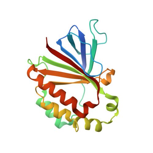
    Chlorite dismutases (Clds) are heme b containing oxidoreductases able to decompose chlorite to chloride and molecular oxygen. This work analyses the impact of the distal, flexible and catalytic arginine on the binding of anionic angulate ligands like nitrite and the substrate chlorite. Dimeric Cld from Cyanothece sp. PCC7425 was used as a model enzyme. We have investigated wild-type CCld having the distal catalytic R127 hydrogen-bonded to glutamine Q74 and variants with R127 (i) being arrested in a salt-bridge with a glutamate (Q74E), (ii) being fully flexible (Q74V) or (iii) substituted by either alanine (R127A) or lysine (R127K). We present the electronic and spectral signatures of the high-spin ferric proteins and the corresponding low-spin nitrite complexes elucidated by UV-visible, circular dichroism and electron paramagnetic resonance spectroscopies. Furthermore, we demonstrate the impact of the dynamics of R127 on the thermal stability of the respective nitrite adducts and present the X-ray crystal structures of the nitrite complexes of wild-type CCld and the variants Q74V, Q74E and R127A. In addition, the molecular dynamics (MD) and the binding modi of nitrite and chlorite to the ferric wild-type enzyme and the mutant proteins and the interaction of the oxoanions with R127 have been analysed by MD simulations. The findings are discussed with respect to the role(s) of R127 in ligand and chlorite binding and substrate degradation.


  • Organizational Affiliation

    BIMEF Laboratory, Department of Chemistry, University of Antwerp, Belgium.


Macromolecules
Find similar proteins by:  (by identity cutoff)  |  3D Structure
Entity ID: 1
MoleculeChains Sequence LengthOrganismDetailsImage
Chlorite dismutase
A, B
188Cyanothece sp. PCC 7425Mutation(s): 1 
Gene Names: Cyan7425_1434
UniProt
Find proteins for B8HNS6 (Cyanothece sp. (strain PCC 7425 / ATCC 29141))
Explore B8HNS6 
Go to UniProtKB:  B8HNS6
Entity Groups  
Sequence Clusters30% Identity50% Identity70% Identity90% Identity95% Identity100% Identity
UniProt GroupB8HNS6
Sequence Annotations
Expand
  • Reference Sequence
Small Molecules
Ligands 6 Unique
IDChains Name / Formula / InChI Key2D Diagram3D Interactions
HEM (Subject of Investigation/LOI)
Query on HEM

Download Ideal Coordinates CCD File 
C [auth A],
I [auth B]
PROTOPORPHYRIN IX CONTAINING FE
C34 H32 Fe N4 O4
KABFMIBPWCXCRK-RGGAHWMASA-L
MES
Query on MES

Download Ideal Coordinates CCD File 
E [auth A],
L [auth B]
2-(N-MORPHOLINO)-ETHANESULFONIC ACID
C6 H13 N O4 S
SXGZJKUKBWWHRA-UHFFFAOYSA-N
PGE
Query on PGE

Download Ideal Coordinates CCD File 
F [auth A],
G [auth A],
K [auth B]
TRIETHYLENE GLYCOL
C6 H14 O4
ZIBGPFATKBEMQZ-UHFFFAOYSA-N
PEG
Query on PEG

Download Ideal Coordinates CCD File 
J [auth B]DI(HYDROXYETHYL)ETHER
C4 H10 O3
MTHSVFCYNBDYFN-UHFFFAOYSA-N
SO4
Query on SO4

Download Ideal Coordinates CCD File 
H [auth A]SULFATE ION
O4 S
QAOWNCQODCNURD-UHFFFAOYSA-L
GOL
Query on GOL

Download Ideal Coordinates CCD File 
D [auth A]GLYCEROL
C3 H8 O3
PEDCQBHIVMGVHV-UHFFFAOYSA-N
Experimental Data & Validation

Experimental Data

  • Method: X-RAY DIFFRACTION
  • Resolution: 2.09 Å
  • R-Value Free: 0.270 
  • R-Value Work: 0.222 
  • R-Value Observed: 0.225 
  • Space Group: P 1
Unit Cell:
Length ( Å )Angle ( ˚ )
a = 48.712α = 102.484
b = 49.607β = 104.838
c = 54.579γ = 105.002
Software Package:
Software NamePurpose
PHENIXrefinement
XDSdata reduction
XSCALEdata scaling
PHASERphasing

Structure Validation

View Full Validation Report


Entry History & Funding Information

Deposition Data


Funding OrganizationLocationGrant Number
Austrian Science FundAustriaP30979

Revision History  (Full details and data files)

  • Version 1.0: 2021-12-22
    Type: Initial release
  • Version 1.1: 2021-12-29
    Changes: Database references
  • Version 1.2: 2024-01-31
    Changes: Data collection, Refinement description