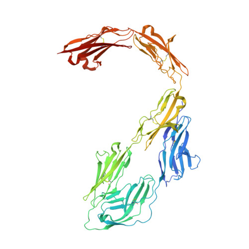
7OL2 | pdb_00007ol2

Crystal structure of mouse contactin 1 immunoglobulin domains


Experimental Data Snapshot

  • Method: X-RAY DIFFRACTION
  • Resolution: 3.89 Å
  • R-Value Free: 
    0.270 (Depositor), 0.270 (DCC) 
  • R-Value Work: 
    0.220 (Depositor), 0.220 (DCC) 
  • R-Value Observed: 
    0.222 (Depositor) 

Starting Model: experimental
View more details

wwPDB Validation   3D Report Full Report


Ligand Structure Quality Assessment 

Created with Raphaël 2.3.0Worse 01 BetterLigand structure goodness of fit to experimental dataBest fitted NAGClick on this verticalbar to view details

This is version 1.2 of the entry. See complete history


Literature

Structural insights into the contactin 1 - neurofascin 155 adhesion complex.

Chataigner, L.M.P.Gogou, C.den Boer, M.A.Frias, C.P.Thies-Weesie, D.M.E.Granneman, J.C.M.Heck, A.J.R.Meijer, D.H.Janssen, B.J.C.

(2022) Nat Commun 13: 6607-6607

  • DOI: https://doi.org/10.1038/s41467-022-34302-9
  • Primary Citation of Related Structures:  
    7OK5, 7OL2, 7OL4

  • PubMed Abstract: 

    Cell-surface expressed contactin 1 and neurofascin 155 control wiring of the nervous system and interact across cells to form and maintain paranodal myelin-axon junctions. The molecular mechanism of contactin 1 - neurofascin 155 adhesion complex formation is unresolved. Crystallographic structures of complexed and individual contactin 1 and neurofascin 155 binding regions presented here, provide a rich picture of how competing and complementary interfaces, post-translational glycosylation, splice differences and structural plasticity enable formation of diverse adhesion sites. Structural, biophysical, and cell-clustering analysis reveal how conserved Ig1-2 interfaces form competing heterophilic contactin 1 - neurofascin 155 and homophilic neurofascin 155 complexes whereas contactin 1 forms low-affinity clusters through interfaces on Ig3-6. The structures explain how the heterophilic Ig1-Ig4 horseshoe's in the contactin 1 - neurofascin 155 complex define the 7.4 nm paranodal spacing and how the remaining six domains enable bridging of distinct intercellular distances.


  • Organizational Affiliation

    Structural Biochemistry, Bijvoet Center for Biomolecular Research, Faculty of Science, Utrecht University, Universiteitsweg 99, 3584 CG, Utrecht, The Netherlands.


Macromolecules
Find similar proteins by:  (by identity cutoff)  |  3D Structure
Entity ID: 1
MoleculeChains Sequence LengthOrganismDetailsImage
Contactin-1
A, B
592Mus musculusMutation(s): 0 
Gene Names: Cntn1
UniProt
Find proteins for P12960 (Mus musculus)
Explore P12960 
Go to UniProtKB:  P12960
Entity Groups  
Sequence Clusters30% Identity50% Identity70% Identity90% Identity95% Identity100% Identity
UniProt GroupP12960
Glycosylation
Glycosylation Sites: 6Go to GlyGen: P12960-1
Sequence Annotations
Expand
  • Reference Sequence
Oligosaccharides

Help

Entity ID: 2
MoleculeChains Length2D Diagram Glycosylation3D Interactions
alpha-D-mannopyranose-(1-3)-[alpha-D-mannopyranose-(1-6)]beta-D-mannopyranose-(1-4)-2-acetamido-2-deoxy-beta-D-glucopyranose-(1-4)-2-acetamido-2-deoxy-beta-D-glucopyranose
C, D
5N-Glycosylation
Glycosylation Resources
GlyTouCan:  G22768VO
GlyCosmos:  G22768VO
GlyGen:  G22768VO
Entity ID: 3
MoleculeChains Length2D Diagram Glycosylation3D Interactions
2-acetamido-2-deoxy-beta-D-glucopyranose-(1-4)-2-acetamido-2-deoxy-beta-D-glucopyranose
E, G, I
2N-Glycosylation
Glycosylation Resources
GlyTouCan:  G42666HT
GlyCosmos:  G42666HT
GlyGen:  G42666HT
Entity ID: 4
MoleculeChains Length2D Diagram Glycosylation3D Interactions
alpha-D-mannopyranose-(1-3)-beta-D-mannopyranose-(1-4)-2-acetamido-2-deoxy-beta-D-glucopyranose-(1-4)-2-acetamido-2-deoxy-beta-D-glucopyranose
F, H
4N-Glycosylation
Glycosylation Resources
GlyTouCan:  G81315DD
GlyCosmos:  G81315DD
GlyGen:  G81315DD
Experimental Data & Validation

Experimental Data

  • Method: X-RAY DIFFRACTION
  • Resolution: 3.89 Å
  • R-Value Free:  0.270 (Depositor), 0.270 (DCC) 
  • R-Value Work:  0.220 (Depositor), 0.220 (DCC) 
  • R-Value Observed: 0.222 (Depositor) 
Space Group: P 21 21 21
Unit Cell:
Length ( Å )Angle ( ˚ )
a = 92.959α = 90
b = 134.162β = 90
c = 181.74γ = 90
Software Package:
Software NamePurpose
PHENIXrefinement
PDB_EXTRACTdata extraction
xia2data reduction
Aimlessdata scaling
PHASERphasing

Structure Validation

View Full Validation Report


Ligand Structure Quality Assessment 

Created with Raphaël 2.3.0Worse 01 BetterLigand structure goodness of fit to experimental dataBest fitted NAGClick on this verticalbar to view details

Entry History & Funding Information

Deposition Data


Funding OrganizationLocationGrant Number
European Research Council (ERC)Netherlands--

Revision History  (Full details and data files)

  • Version 1.0: 2022-12-14
    Type: Initial release
  • Version 1.1: 2024-01-31
    Changes: Data collection, Refinement description
  • Version 1.2: 2024-11-13
    Changes: Structure summary