7OH6

Cryo-EM structure of Drs2p-Cdc50p in the [PS]E2-AlFx state


Experimental Data Snapshot

  • Method: ELECTRON MICROSCOPY
  • Resolution: 3.00 Å
  • Aggregation State: PARTICLE 
  • Reconstruction Method: SINGLE PARTICLE 

wwPDB Validation   3D Report Full Report


This is version 1.0 of the entry. See complete history


Literature

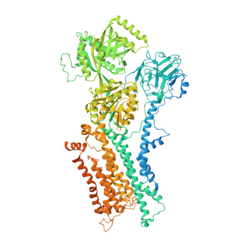
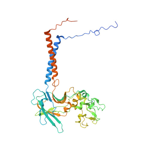
Structural basis of substrate-independent phosphorylation in a P4-ATPase lipid flippase

Timcenko, M.Dieudonne, T.Montigny, C.Boesen, T.Lyons, J.A.Lenoir, G.Nissen, P.

(2021) J Mol Biol : 167062

  • DOI: https://doi.org/10.1016/j.jmb.2021.167062
  • Primary Citation of Related Structures:  
    7OH4, 7OH5, 7OH6, 7OH7

  • PubMed Abstract: 

    P4-ATPases define a eukaryotic subfamily of the P-type ATPases, and are responsible for the transverse flip of specific lipids from the extracellular or luminal leaflet to the cytosolic leaflet of cell membranes. The enzymatic cycle of P-type ATPases is divided into autophosphorylation and dephosphorylation half-reactions. Unlike most other P-type ATPases, P4-ATPases transport their substrate during dephosphorylation only, i.e. the phosphorylation half-reaction is not associated with transport. To study the structural basis of the distinct mechanisms of P4-ATPases, we have determined cryo-EM structures of Drs2p-Cdc50p from Saccharomyces cerevisiae covering multiple intermediates of the cycle. We identify several structural motifs specific to Drs2p and P4-ATPases in general that decrease movements and flexibility of domains as compared to other P-type ATPases such as Na + /K + -ATPase or Ca 2+ -ATPase. These motifs include the linkers that connect the transmembrane region to the actuator (A) domain, which is responsible for dephosphorylation. Additionally, mutation of Tyr380, which interacts with conserved Asp340 of the distinct DGET dephosphorylation loop of P4-ATPases, highlights a functional role of these P4-ATPase specific motifs in the A-domain. Finally, the transmembrane (TM) domain, responsible for transport, also undergoes less extensive conformational changes, which is ensured both by a longer segment connecting TM helix 4 with the phosphorylation site, and possible stabilization by the auxiliary subunit Cdc50p. Collectively these adaptions in P4-ATPases are responsible for phosphorylation becoming transport-independent.


  • Organizational Affiliation

    Danish Research Institute of Translational Neuroscience - DANDRITE, Nordic EMBL Partnerhip for Molecular Medicine, Aarhus University, Dept. Molecular Biology and Genetics, Gustav Wieds Vej 10C, DK - 8000 Aarhus C, Denmark.


Macromolecules
Find similar proteins by:  (by identity cutoff)  |  3D Structure
Entity ID: 1
MoleculeChains Sequence LengthOrganismDetailsImage
Probable phospholipid-transporting ATPase DRS2,Probable phospholipid-transporting ATPase DRS21,465Saccharomyces cerevisiae S288CMutation(s): 0 
Gene Names: DRS2YAL026CFUN38
EC: 7.6.2.1
Membrane Entity: Yes 
UniProt
Find proteins for P39524 (Saccharomyces cerevisiae (strain ATCC 204508 / S288c))
Explore P39524 
Go to UniProtKB:  P39524
Entity Groups  
Sequence Clusters30% Identity50% Identity70% Identity90% Identity95% Identity100% Identity
UniProt GroupP39524
Sequence Annotations
Expand
  • Reference Sequence
Find similar proteins by:  (by identity cutoff)  |  3D Structure
Entity ID: 2
MoleculeChains Sequence LengthOrganismDetailsImage
Cell division control protein 50B [auth C]413Saccharomyces cerevisiae S288CMutation(s): 0 
Gene Names: CDC50YCR094WYCR94W
Membrane Entity: Yes 
UniProt
Find proteins for P25656 (Saccharomyces cerevisiae (strain ATCC 204508 / S288c))
Explore P25656 
Go to UniProtKB:  P25656
Entity Groups  
Sequence Clusters30% Identity50% Identity70% Identity90% Identity95% Identity100% Identity
UniProt GroupP25656
Sequence Annotations
Expand
  • Reference Sequence
Oligosaccharides

Help

Entity ID: 3
MoleculeChains Length2D Diagram Glycosylation3D Interactions
2-acetamido-2-deoxy-beta-D-glucopyranose-(1-4)-2-acetamido-2-deoxy-beta-D-glucopyranoseC [auth B]2N-Glycosylation
Glycosylation Resources
GlyTouCan:  G42666HT
GlyCosmos:  G42666HT
GlyGen:  G42666HT
Entity ID: 4
MoleculeChains Length2D Diagram Glycosylation3D Interactions
beta-D-mannopyranose-(1-4)-2-acetamido-2-deoxy-beta-D-glucopyranose-(1-4)-2-acetamido-2-deoxy-beta-D-glucopyranose
D
3N/AN-Glycosylation
Small Molecules
Ligands 5 Unique
IDChains Name / Formula / InChI Key2D Diagram3D Interactions
2Y5
Query on 2Y5

Download Ideal Coordinates CCD File 
E [auth A](2R)-1-{[(R)-hydroxy{[(1R,2R,3R,4R,5S,6R)-2,3,5,6-tetrahydroxy-4-(phosphonooxy)cyclohexyl]oxy}phosphoryl]oxy}-3-(octadecanoyloxy)propan-2-yl (5Z,8Z,11Z,14Z)-icosa-5,8,11,14-tetraenoate
C47 H84 O16 P2
ROKMWIUSHIHOGI-BWTMTLBLSA-N
Q3G
Query on Q3G

Download Ideal Coordinates CCD File 
H [auth A]O-[(R)-[(2S)-2-(hexadecanoyloxy)-3-(octadecanoyloxy)propoxy](hydroxy)phosphoryl]-D-serine
C40 H78 N O10 P
WZFUPCSEUKNOBF-PQQNNWGCSA-N
NAG
Query on NAG

Download Ideal Coordinates CCD File 
I [auth C]2-acetamido-2-deoxy-beta-D-glucopyranose
C8 H15 N O6
OVRNDRQMDRJTHS-FMDGEEDCSA-N
ALF
Query on ALF

Download Ideal Coordinates CCD File 
F [auth A]TETRAFLUOROALUMINATE ION
Al F4
UYOMQIYKOOHAMK-UHFFFAOYSA-J
MG
Query on MG

Download Ideal Coordinates CCD File 
G [auth A]MAGNESIUM ION
Mg
JLVVSXFLKOJNIY-UHFFFAOYSA-N
Experimental Data & Validation

Experimental Data

  • Method: ELECTRON MICROSCOPY
  • Resolution: 3.00 Å
  • Aggregation State: PARTICLE 
  • Reconstruction Method: SINGLE PARTICLE 
EM Software:
TaskSoftware PackageVersion
RECONSTRUCTIONcryoSPARC2
MODEL REFINEMENTPHENIX1.18rc7-3834

Structure Validation

View Full Validation Report


Entry History & Funding Information

Deposition Data


Funding OrganizationLocationGrant Number
LundbeckfondenDenmark--
Novo Nordisk FoundationDenmark--
Boehringer Ingelheim Fonds (BIF)Germany--

Revision History  (Full details and data files)

  • Version 1.0: 2021-06-09
    Type: Initial release