7O82 | pdb_00007o82

The L-arginine/agmatine antiporter from E. coli at 1.7 A resolution


Experimental Data Snapshot

  • Method: X-RAY DIFFRACTION
  • Resolution: 1.69 Å
  • R-Value Free: 
    0.203 (Depositor), 0.210 (DCC) 
  • R-Value Work: 
    0.195 (Depositor), 0.200 (DCC) 
  • R-Value Observed: 
    0.195 (Depositor) 

Starting Model: experimental
View more details

wwPDB Validation 3D Report Full Report

Validation slider image for 7O82

Ligand Structure Quality Assessment 


This is version 1.1 of the entry. See complete history

Literature

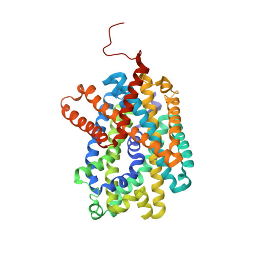
High-resolution structure of the amino acid transporter AdiC reveals insights into the role of water molecules and networks in oligomerization and substrate binding.

Ilgu, H.Jeckelmann, J.M.Kalbermatter, D.Ucurum, Z.Lemmin, T.Fotiadis, D.

(2021) BMC Biol 19: 179-179

  • DOI: https://doi.org/10.1186/s12915-021-01102-4
  • Primary Citation Related Structures: 
    7O82

  • PubMed Abstract: 

    The L-arginine/agmatine transporter AdiC is part of the arginine-dependent extreme acid resistance system of the bacterium Escherichia coli and its pathogenic varieties such as strain E. coli O157:H7. At the present time, there is a lack of knowledge concerning the role of water molecules and networks for the structure and function of AdiC, and solute transporters in general. The structure of the L-arginine/agmatine transporter AdiC was determined at 1.7 Å resolution by X-ray crystallography. This high resolution allowed for the identification of numerous water molecules buried in the structure. In combination with molecular dynamics (MD) simulations, we demonstrate that water molecules play an important role for stabilizing the protein and key residues, and act as placeholders for atoms of the AdiC substrates L-arginine and agmatine. MD simulations unveiled flexibility and restrained mobility of gating residues W202 and W293, respectively. Furthermore, a water-filled cavity was identified at the dimer interface of AdiC. The two monomers formed bridging interactions through water-mediated hydrogen bonds. The accessibility and presence of water molecules in this cavity was confirmed with MD simulations. Point mutations disrupting the interfacial water network validated the importance of water molecules for dimer stabilization. This work gives new insights into the role and importance of water molecules in the L-arginine/agmatine transporter AdiC for protein stabilization and substrate-binding site shaping and as placeholders of substrate atoms. Furthermore, and based on the observed flexibility and restrained mobility of gating residues, a mechanistic role of the gate flexibility in the transport cycle was proposed. Finally, we identified a water-filled cavity at the dimeric interface that contributes to the stability of the amino acid transporter oligomer.


  • Organizational Affiliation
    • Institute of Biochemistry and Molecular Medicine, and Swiss National Centre of Competence in Research (NCCR) TransCure, University of Bern, CH-3012, Bern, Switzerland.

Macromolecule Content 

  • Total Structure Weight: 97.76 kDa 
  • Atom Count: 7,053 
  • Modeled Residue Count: 873 
  • Deposited Residue Count: 906 
  • Unique protein chains: 1

Macromolecules

Find similar proteins by:|  3D Structure
Entity ID: 1
MoleculeChains  Sequence LengthOrganismDetailsImage
Arginine/agmatine antiporter
A, B
453Escherichia coli O157:H7Mutation(s): 0 
Gene Names: adiCZ5717ECs5097
Membrane Entity: Yes 
UniProt
Find proteins for P60061 (Escherichia coli (strain K12))
Explore P60061 
Go to UniProtKB:  P60061
Entity Groups
Sequence Clusters30% Identity50% Identity70% Identity90% Identity95% Identity100% Identity
UniProt GroupP60061
Sequence Annotations
Expand
Reference Sequence

Small Molecules

Ligands 5 Unique
IDChains Name / Formula / InChI Key2D Diagram3D Interactions
BNG

Query on BNG


Download:Ideal Coordinates CCD File
C [auth A],
H [auth B],
I [auth B]
nonyl beta-D-glucopyranoside
C15 H30 O6
QFAPUKLCALRPLH-UXXRCYHCSA-N
D10

Query on D10


Download:Ideal Coordinates CCD File
F [auth A],
P [auth B]
DECANE
C10 H22
DIOQZVSQGTUSAI-UHFFFAOYSA-N
TRS

Query on TRS


Download:Ideal Coordinates CCD File
D [auth A],
J [auth B]
2-AMINO-2-HYDROXYMETHYL-PROPANE-1,3-DIOL
C4 H12 N O3
LENZDBCJOHFCAS-UHFFFAOYSA-O
OCT

Query on OCT


Download:Ideal Coordinates CCD File
G [auth A]N-OCTANE
C8 H18
TVMXDCGIABBOFY-UHFFFAOYSA-N
HEX

Query on HEX


Download:Ideal Coordinates CCD File
E [auth A]
K [auth B]
L [auth B]
M [auth B]
N [auth B]
E [auth A],
K [auth B],
L [auth B],
M [auth B],
N [auth B],
O [auth B]
HEXANE
C6 H14
VLKZOEOYAKHREP-UHFFFAOYSA-N

Experimental Data & Validation

Experimental Data

  • Method: X-RAY DIFFRACTION
  • Resolution: 1.69 Å
  • R-Value Free:  0.203 (Depositor), 0.210 (DCC) 
  • R-Value Work:  0.195 (Depositor), 0.200 (DCC) 
  • R-Value Observed: 0.195 (Depositor) 
Space Group: P 21 21 2
Unit Cell:
Length ( Å )Angle ( ˚ )
a = 104.708α = 90
b = 175.592β = 90
c = 73.275γ = 90
Software Package:
Software NamePurpose
Aimlessdata scaling
PHASERphasing
PHENIXrefinement
PDB_EXTRACTdata extraction
XDSdata reduction

Structure Validation

View Full Validation Report


Ligand Structure Quality Assessment 


Entry History 

& Funding Information

Deposition Data


Funding OrganizationLocationGrant Number
Swiss National Science FoundationSwitzerland310030_184980

Revision History  (Full details and data files)

  • Version 1.0: 2021-09-08
    Type: Initial release
  • Version 1.1: 2024-01-31
    Changes: Data collection, Refinement description