7NWQ

A carbohydrate binding module family 9 (CBM9) from Caldicellulosiruptor kristjanssonii in complex with cellotriose


Experimental Data Snapshot

  • Method: X-RAY DIFFRACTION
  • Resolution: 2.23 Å
  • R-Value Free: 0.226 
  • R-Value Work: 0.185 

wwPDB Validation   3D Report Full Report


This is version 1.2 of the entry. See complete history


Literature

Structural and Functional Analysis of a Multimodular Hyperthermostable Xylanase-Glucuronoyl Esterase from Caldicellulosiruptor kristjansonii .

Krska, D.Mazurkewich, S.Brown, H.A.Theibich, Y.Poulsen, J.N.Morris, A.L.Koropatkin, N.M.Lo Leggio, L.Larsbrink, J.

(2021) Biochemistry 60: 2206-2220

  • DOI: https://doi.org/10.1021/acs.biochem.1c00305
  • Primary Citation of Related Structures:  
    7NN3, 7NWN, 7NWO, 7NWP, 7NWQ

  • PubMed Abstract: 

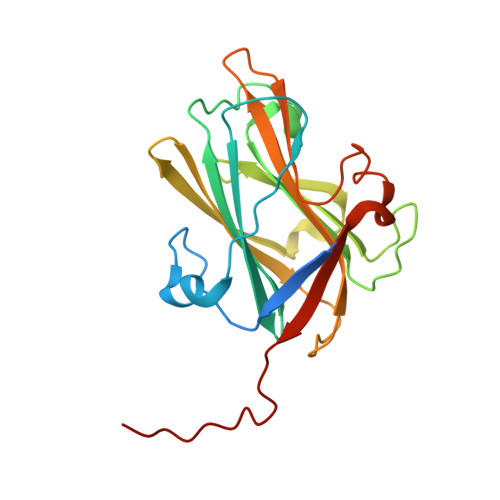
    The hyperthermophilic bacterium Caldicellulosiruptor kristjansonii encodes an unusual enzyme, Ck Xyn10C-GE15A, which incorporates two catalytic domains, a xylanase and a glucuronoyl esterase, and five carbohydrate-binding modules (CBMs) from families 9 and 22. The xylanase and glucuronoyl esterase catalytic domains were recently biochemically characterized, as was the ability of the individual CBMs to bind insoluble polysaccharides. Here, we further probed the abilities of the different CBMs from Ck Xyn10C-GE15A to bind to soluble poly- and oligosaccharides using affinity gel electrophoresis, isothermal titration calorimetry, and differential scanning fluorimetry. The results revealed additional binding properties of the proteins compared to the former studies on insoluble polysaccharides. Collectively, the results show that all five CBMs have their own distinct binding preferences and appear to complement each other and the catalytic domains in targeting complex cell wall polysaccharides. Additionally, through renewed efforts, we have achieved partial structural characterization of this complex multidomain protein. We have determined the structures of the third CBM9 domain (CBM9.3) and the glucuronoyl esterase (GE15A) by X-ray crystallography. CBM9.3 is the second CBM9 structure determined to date and was shown to bind oligosaccharide ligands at the same site but in a different binding mode compared to that of the previously determined CBM9 structure from Thermotoga maritima . GE15A represents a unique intermediate between reported fungal and bacterial glucuronoyl esterase structures as it lacks two inserted loop regions typical of bacterial enzymes and a third loop has an atypical structure. We also report small-angle X-ray scattering measurements of the N-terminal CBM22.1-CBM22.2-Xyn10C construct, indicating a compact arrangement at room temperature.


  • Organizational Affiliation

    Division of Industrial Biotechnology, Department of Biology and Biological Engineering, Chalmers University of Technology, SE-412 96 Gothenburg, Sweden.


Macromolecules
Find similar proteins by:  (by identity cutoff)  |  3D Structure
Entity ID: 1
MoleculeChains Sequence LengthOrganismDetailsImage
Beta-xylanaseA [auth AAA]215Caldicellulosiruptor acetigenus I77R1BMutation(s): 0 
Gene Names: Calkr_2245
EC: 3.2.1.8
UniProt
Find proteins for E4S6E9 (Caldicellulosiruptor acetigenus (strain ATCC 700853 / DSM 12137 / I77R1B))
Explore E4S6E9 
Go to UniProtKB:  E4S6E9
Entity Groups  
Sequence Clusters30% Identity50% Identity70% Identity90% Identity95% Identity100% Identity
UniProt GroupE4S6E9
Sequence Annotations
Expand
  • Reference Sequence
Oligosaccharides

Help

Entity ID: 2
MoleculeChains Length2D Diagram Glycosylation3D Interactions
beta-D-glucopyranose-(1-4)-beta-D-glucopyranose-(1-4)-beta-D-glucopyranoseB [auth BBB]3N/A
Glycosylation Resources
GlyTouCan:  G14338VK
GlyCosmos:  G14338VK
GlyGen:  G14338VK
Small Molecules
Ligands 5 Unique
IDChains Name / Formula / InChI Key2D Diagram3D Interactions
1PE
Query on 1PE

Download Ideal Coordinates CCD File 
W [auth AAA]PENTAETHYLENE GLYCOL
C10 H22 O6
JLFNLZLINWHATN-UHFFFAOYSA-N
PGE
Query on PGE

Download Ideal Coordinates CCD File 
V [auth AAA],
X [auth AAA]
TRIETHYLENE GLYCOL
C6 H14 O4
ZIBGPFATKBEMQZ-UHFFFAOYSA-N
PEG
Query on PEG

Download Ideal Coordinates CCD File 
C [auth AAA]
D [auth AAA]
E [auth AAA]
F [auth AAA]
G [auth AAA]
C [auth AAA],
D [auth AAA],
E [auth AAA],
F [auth AAA],
G [auth AAA],
H [auth AAA],
I [auth AAA]
DI(HYDROXYETHYL)ETHER
C4 H10 O3
MTHSVFCYNBDYFN-UHFFFAOYSA-N
EDO
Query on EDO

Download Ideal Coordinates CCD File 
J [auth AAA]
K [auth AAA]
L [auth AAA]
M [auth AAA]
N [auth AAA]
J [auth AAA],
K [auth AAA],
L [auth AAA],
M [auth AAA],
N [auth AAA],
O [auth AAA],
P [auth AAA],
Q [auth AAA],
R [auth AAA]
1,2-ETHANEDIOL
C2 H6 O2
LYCAIKOWRPUZTN-UHFFFAOYSA-N
CA
Query on CA

Download Ideal Coordinates CCD File 
S [auth AAA],
T [auth AAA],
U [auth AAA]
CALCIUM ION
Ca
BHPQYMZQTOCNFJ-UHFFFAOYSA-N
Biologically Interesting Molecules (External Reference) 1 Unique
Experimental Data & Validation

Experimental Data

  • Method: X-RAY DIFFRACTION
  • Resolution: 2.23 Å
  • R-Value Free: 0.226 
  • R-Value Work: 0.185 
  • Space Group: I 4 3 2
Unit Cell:
Length ( Å )Angle ( ˚ )
a = 172.49α = 90
b = 172.49β = 90
c = 172.49γ = 90
Software Package:
Software NamePurpose
REFMACrefinement
PHENIXphasing

Structure Validation

View Full Validation Report


Entry History & Funding Information

Deposition Data


Funding OrganizationLocationGrant Number
Swedish Research CouncilSweden2016-01065
Swedish Energy AgencySweden2016-011207
Novo Nordisk FoundationDenmarkNNF17OC0027698
Knut and Alice Wallenberg FoundationSweden--

Revision History  (Full details and data files)

  • Version 1.0: 2021-07-07
    Type: Initial release
  • Version 1.1: 2021-07-21
    Changes: Database references
  • Version 1.2: 2024-01-31
    Changes: Data collection, Database references, Derived calculations, Refinement description