7NTU

X-ray structure of the complex between human alpha thrombin and two duplex/quadruplex aptamers: NU172 and HD22_27mer


Experimental Data Snapshot

  • Method: X-RAY DIFFRACTION
  • Resolution: 3.10 Å
  • R-Value Free: 0.257 
  • R-Value Work: 0.200 
  • R-Value Observed: 0.203 

wwPDB Validation   3D Report Full Report


Ligand Structure Quality Assessment 


This is version 1.2 of the entry. See complete history


Literature

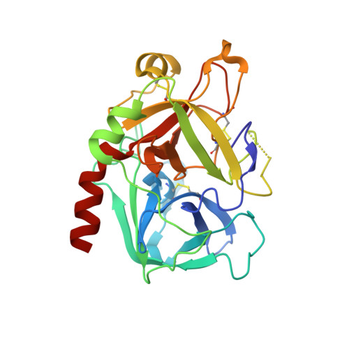
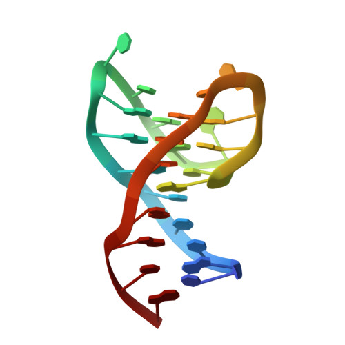
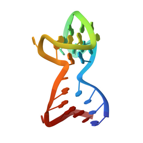
Structural and functional analysis of the simultaneous binding of two duplex/quadruplex aptamers to human alpha-thrombin.

Troisi, R.Balasco, N.Santamaria, A.Vitagliano, L.Sica, F.

(2021) Int J Biol Macromol 181: 858-867

  • DOI: https://doi.org/10.1016/j.ijbiomac.2021.04.076
  • Primary Citation of Related Structures:  
    7NTU

  • PubMed Abstract: 

    The long-range communication between the two exosites of human α-thrombin (thrombin) tightly modulates the protein-effector interactions. Duplex/quadruplex aptamers represent an emerging class of very effective binders of thrombin. Among them, NU172 and HD22 aptamers are at the forefront of exosite I and II recognition, respectively. The present study investigates the simultaneous binding of these two aptamers by combining a structural and dynamics approach. The crystal structure of the ternary complex formed by the thrombin with NU172 and HD22_27mer provides a detailed view of the simultaneous binding of these aptamers to the protein, inspiring the design of novel bivalent thrombin inhibitors. The crystal structure represents the starting model for molecular dynamics studies, which point out the cooperation between the binding at the two exosites. In particular, the binding of an aptamer to its exosite reduces the intrinsic flexibility of the other exosite, that preferentially assumes conformations similar to those observed in the bound state, suggesting a predisposition to interact with the other aptamer. This behaviour is reflected in a significant increase of the anticoagulant activity of NU172 when the inactive HD22_27mer is bound to exosite II, providing a clear evidence of the synergic action of the two aptamers.


  • Organizational Affiliation

    Department of Chemical Sciences, University of Naples Federico II, Complesso Universitario di Monte Sant'Angelo, Via Cintia, I-80126 Naples, Italy.


Macromolecules

Find similar proteins by:  (by identity cutoff)  |  3D Structure
Entity ID: 1
MoleculeChains Sequence LengthOrganismDetailsImage
Prothrombin
A, C
36Homo sapiensMutation(s): 0 
EC: 3.4.21.5
UniProt & NIH Common Fund Data Resources
Find proteins for P00734 (Homo sapiens)
Explore P00734 
Go to UniProtKB:  P00734
PHAROS:  P00734
GTEx:  ENSG00000180210 
Entity Groups  
Sequence Clusters30% Identity50% Identity70% Identity90% Identity95% Identity100% Identity
UniProt GroupP00734
Sequence Annotations
Expand
  • Reference Sequence
Find similar proteins by:  (by identity cutoff)  |  3D Structure
Entity ID: 2
MoleculeChains Sequence LengthOrganismDetailsImage
Prothrombin
B, D
259Homo sapiensMutation(s): 0 
EC: 3.4.21.5
UniProt & NIH Common Fund Data Resources
Find proteins for P00734 (Homo sapiens)
Explore P00734 
Go to UniProtKB:  P00734
PHAROS:  P00734
GTEx:  ENSG00000180210 
Entity Groups  
Sequence Clusters30% Identity50% Identity70% Identity90% Identity95% Identity100% Identity
UniProt GroupP00734
Sequence Annotations
Expand
  • Reference Sequence
Find similar nucleic acids by:  (by identity cutoff)  |  3D Structure
Entity ID: 3
MoleculeChains LengthOrganismImage
NU172
E, G
26synthetic construct
Sequence Annotations
Expand
  • Reference Sequence
Find similar nucleic acids by:  (by identity cutoff)  |  3D Structure
Entity ID: 4
MoleculeChains LengthOrganismImage
HD22_27merF,
H [auth I]
27synthetic construct
Sequence Annotations
Expand
  • Reference Sequence
Small Molecules
Ligands 3 Unique
IDChains Name / Formula / InChI Key2D Diagram3D Interactions
0G6
Query on 0G6

Download Ideal Coordinates CCD File 
I [auth B],
K [auth D]
D-phenylalanyl-N-[(2S,3S)-6-{[amino(iminio)methyl]amino}-1-chloro-2-hydroxyhexan-3-yl]-L-prolinamide
C21 H34 Cl N6 O3
DVFLYEYCMMLBTQ-VSZNYVQBSA-O
NAG
Query on NAG

Download Ideal Coordinates CCD File 
J [auth B],
L [auth D]
2-acetamido-2-deoxy-beta-D-glucopyranose
C8 H15 N O6
OVRNDRQMDRJTHS-FMDGEEDCSA-N
NA
Query on NA

Download Ideal Coordinates CCD File 
M [auth E],
N [auth F],
O [auth G],
P [auth I]
SODIUM ION
Na
FKNQFGJONOIPTF-UHFFFAOYSA-N
Biologically Interesting Molecules (External Reference) 1 Unique
Experimental Data & Validation

Experimental Data

  • Method: X-RAY DIFFRACTION
  • Resolution: 3.10 Å
  • R-Value Free: 0.257 
  • R-Value Work: 0.200 
  • R-Value Observed: 0.203 
  • Space Group: P 1 21 1
Unit Cell:
Length ( Å )Angle ( ˚ )
a = 84.927α = 90
b = 77.616β = 103.47
c = 95.408γ = 90
Software Package:
Software NamePurpose
REFMACrefinement
PDB_EXTRACTdata extraction
HKL-2000data reduction
HKL-2000data scaling
PHASERphasing

Structure Validation

View Full Validation Report


Ligand Structure Quality Assessment 


Entry History 

Deposition Data

Revision History  (Full details and data files)

  • Version 1.0: 2021-04-28
    Type: Initial release
  • Version 1.1: 2021-05-05
    Changes: Database references
  • Version 1.2: 2024-01-31
    Changes: Data collection, Database references, Refinement description