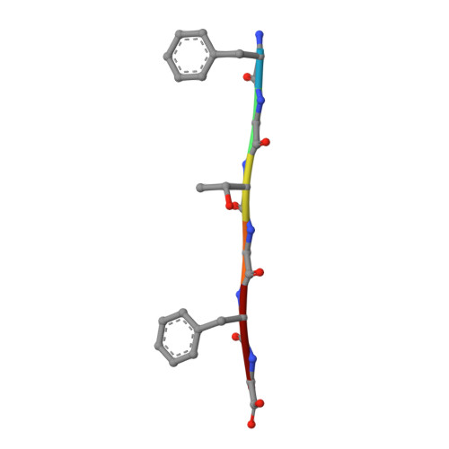
7N8R

FGTGFG segment from the Nucleoporin p54, residues 63-68


Experimental Data Snapshot

  • Method: X-RAY DIFFRACTION
  • Resolution: 1.20 Å
  • R-Value Free: 0.174 
  • R-Value Work: 0.143 
  • R-Value Observed: 0.146 

wwPDB Validation   3D Report Full Report


This is version 1.1 of the entry. See complete history


Literature

Extended beta-Strands Contribute to Reversible Amyloid Formation.

Murray, K.A.Evans, D.Hughes, M.P.Sawaya, M.R.Hu, C.J.Houk, K.N.Eisenberg, D.

(2022) ACS Nano 16: 2154-2163

  • DOI: https://doi.org/10.1021/acsnano.1c08043
  • Primary Citation of Related Structures:  
    7N8R

  • PubMed Abstract: 

    The assembly of proteins into fibrillar amyloid structures was once considered to be pathologic and essentially irreversible. Recent studies reveal amyloid-like structures that form reversibly, derived from protein low-complexity domains which function in cellular metabolism. Here, by comparing atomic-level structures of reversible and irreversible amyloid fibrils, we find that the β-sheets of reversible fibrils are enriched in flattened (as opposed to pleated) β-sheets formed by stacking of extended β-strands. Quantum mechanical calculations show that glycine residues favor extended β-strands which may be stabilized by intraresidue interactions between the amide proton and the carbonyl oxygen, known as C5 hydrogen-bonds. Larger residue side chains favor shorter strands and pleated sheets. These findings highlight a structural element that may regulate reversible amyloid assembly.


Macromolecules

Find similar proteins by:  Sequence   |   3D Structure  

Entity ID: 1
MoleculeChains Sequence LengthOrganismDetailsImage
FGTGFG segment from the Nucleoporin p54, residues 63-68
A, B
6Homo sapiensMutation(s): 0 
UniProt & NIH Common Fund Data Resources
Find proteins for Q7Z3B4 (Homo sapiens)
Explore Q7Z3B4 
Go to UniProtKB:  Q7Z3B4
PHAROS:  Q7Z3B4
GTEx:  ENSG00000138750 
Entity Groups  
UniProt GroupQ7Z3B4
Sequence Annotations
Expand
  • Reference Sequence
Small Molecules
Ligands 1 Unique
IDChains Name / Formula / InChI Key2D Diagram3D Interactions
TFA
Query on TFA

Download Ideal Coordinates CCD File 
C [auth B]trifluoroacetic acid
C2 H F3 O2
DTQVDTLACAAQTR-UHFFFAOYSA-N
Experimental Data & Validation

Experimental Data

  • Method: X-RAY DIFFRACTION
  • Resolution: 1.20 Å
  • R-Value Free: 0.174 
  • R-Value Work: 0.143 
  • R-Value Observed: 0.146 
  • Space Group: P 1 21 1
Unit Cell:
Length ( Å )Angle ( ˚ )
a = 15.71α = 90
b = 9.4β = 108.52
c = 23.26γ = 90
Software Package:
Software NamePurpose
XDSdata reduction
XSCALEdata scaling
PHASERphasing
REFMACrefinement
PDB_EXTRACTdata extraction

Structure Validation

View Full Validation Report


Entry History & Funding Information

Deposition Data


Funding OrganizationLocationGrant Number
National Institutes of Health/National Institute of General Medical Sciences (NIH/NIGMS)United StatesGM08042

Revision History  (Full details and data files)

  • Version 1.0: 2022-02-16
    Type: Initial release
  • Version 1.1: 2022-03-09
    Changes: Database references