7N8H | pdb_00007n8h

SARS-CoV-2 S (B.1.429 / epsilon variant) + S2M11 + S2L20 Global Refinement


Experimental Data Snapshot

  • Method: ELECTRON MICROSCOPY
  • Resolution: 2.30 Å
  • Aggregation State: PARTICLE 
  • Reconstruction Method: SINGLE PARTICLE 

wwPDB Validation 3D Report Full Report

Validation slider image for 7N8H

This is version 1.2 of the entry. See complete history

Literature

SARS-CoV-2 immune evasion by the B.1.427/B.1.429 variant of concern.

McCallum, M.Bassi, J.De Marco, A.Chen, A.Walls, A.C.Di Iulio, J.Tortorici, M.A.Navarro, M.J.Silacci-Fregni, C.Saliba, C.Sprouse, K.R.Agostini, M.Pinto, D.Culap, K.Bianchi, S.Jaconi, S.Cameroni, E.Bowen, J.E.Tilles, S.W.Pizzuto, M.S.Guastalla, S.B.Bona, G.Pellanda, A.F.Garzoni, C.Van Voorhis, W.C.Rosen, L.E.Snell, G.Telenti, A.Virgin, H.W.Piccoli, L.Corti, D.Veesler, D.

(2021) Science 373: 648-654

  • DOI: https://doi.org/10.1126/science.abi7994
  • Primary Citation Related Structures: 
    7N8H, 7N8I

  • PubMed Abstract: 

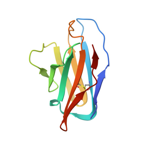
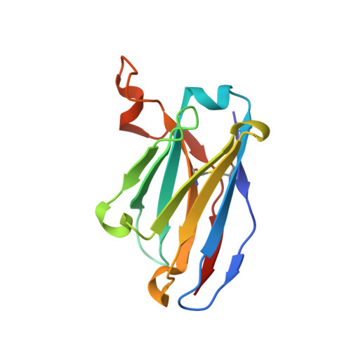
    A novel variant of concern (VOC) named CAL.20C (B.1.427/B.1.429), which was originally detected in California, carries spike glycoprotein mutations S13I in the signal peptide, W152C in the N-terminal domain (NTD), and L452R in the receptor-binding domain (RBD). Plasma from individuals vaccinated with a Wuhan-1 isolate-based messenger RNA vaccine or from convalescent individuals exhibited neutralizing titers that were reduced 2- to 3.5-fold against the B.1.427/B.1.429 variant relative to wild-type pseudoviruses. The L452R mutation reduced neutralizing activity in 14 of 34 RBD-specific monoclonal antibodies (mAbs). The S13I and W152C mutations resulted in total loss of neutralization for 10 of 10 NTD-specific mAbs because the NTD antigenic supersite was remodeled by a shift of the signal peptide cleavage site and the formation of a new disulfide bond, as revealed by mass spectrometry and structural studies.


  • Organizational Affiliation
    • Department of Biochemistry, University of Washington, Seattle, WA 98195, USA.

Macromolecule Content 

  • Total Structure Weight: 587.38 kDa 
  • Atom Count: 32,921 
  • Modeled Residue Count: 4,356 
  • Deposited Residue Count: 5,190 
  • Unique protein chains: 5

Macromolecules

Find similar proteins by:|  3D Structure
Entity ID: 1
MoleculeChains  Sequence LengthOrganismDetailsImage
Spike glycoprotein
A, F, K
1,277Severe acute respiratory syndrome coronavirus 2Mutation(s): 0 
Gene Names: S2
UniProt
Find proteins for P0DTC2 (Severe acute respiratory syndrome coronavirus 2)
Explore P0DTC2 
Go to UniProtKB:  P0DTC2
Entity Groups
Sequence Clusters30% Identity50% Identity70% Identity90% Identity95% Identity100% Identity
UniProt GroupP0DTC2
Glycosylation
Glycosylation Sites: 16Go to GlyGen: P0DTC2-1
Sequence Annotations
Expand
Reference Sequence
Find similar proteins by:|  3D Structure
Entity ID: 2
MoleculeChains  Sequence LengthOrganismDetailsImage
S2M11 Fab Light Chain variable regionB [auth D],
G,
L
104Homo sapiensMutation(s): 0 
Find similar proteins by:|  3D Structure
Entity ID: 3
MoleculeChains  Sequence LengthOrganismDetailsImage
S2M11 Fab Heavy Chain variable regionC [auth E],
H,
M
122Homo sapiensMutation(s): 0 
Find similar proteins by:|  3D Structure
Entity ID: 4
MoleculeChains  Sequence LengthOrganismDetailsImage
S2L20 Fab Light Chain variable regionD [auth B],
I,
N
106Homo sapiensMutation(s): 0 
Find similar proteins by:|  3D Structure
Entity ID: 5
MoleculeChains  Sequence LengthOrganismDetailsImage
S2L20 Fab Heavy Chain variable regionE [auth C],
J,
O
121Homo sapiensMutation(s): 0 

Oligosaccharides

Help  
Entity ID: 6
MoleculeChains Length2D Diagram GlycosylationD Interactions
2-acetamido-2-deoxy-beta-D-glucopyranose-(1-4)-[alpha-L-fucopyranose-(1-6)]2-acetamido-2-deoxy-beta-D-glucopyranose
P, S, V
3N-Glycosylation
Glycosylation Resources
GlyTouCan: G21290RB
GlyCosmos: G21290RB
GlyGen: G21290RB
Entity ID: 7
MoleculeChains Length2D Diagram GlycosylationD Interactions
2-acetamido-2-deoxy-beta-D-glucopyranose-(1-4)-2-acetamido-2-deoxy-beta-D-glucopyranose
Q, R, T, U, W
Q, R, T, U, W, X
2N-Glycosylation
Glycosylation Resources
GlyTouCan: G42666HT
GlyCosmos: G42666HT
GlyGen: G42666HT

Small Molecules

Ligands 1 Unique
IDChains Name / Formula / InChI Key2D Diagram3D Interactions
NAG
(Subject of Investigation/LOI)

Query on NAG


Download:Ideal Coordinates CCD File
AA [auth A]
AB [auth K]
BA [auth A]
BB [auth K]
CA [auth A]
AA [auth A],
AB [auth K],
BA [auth A],
BB [auth K],
CA [auth A],
CB [auth K],
DA [auth A],
DB [auth K],
EA [auth A],
EB [auth K],
FA [auth A],
FB [auth K],
GA [auth A],
GB [auth K],
HA [auth A],
HB [auth K],
IA [auth A],
IB [auth K],
JA [auth A],
JB [auth K],
KA [auth A],
KB [auth K],
LA [auth F],
MA [auth F],
NA [auth F],
OA [auth F],
PA [auth F],
QA [auth F],
RA [auth F],
SA [auth F],
TA [auth F],
UA [auth F],
VA [auth F],
WA [auth F],
XA [auth F],
Y [auth A],
YA [auth K],
Z [auth A],
ZA [auth K]
2-acetamido-2-deoxy-beta-D-glucopyranose
C8 H15 N O6
OVRNDRQMDRJTHS-FMDGEEDCSA-N

Experimental Data & Validation

Experimental Data

  • Method: ELECTRON MICROSCOPY
  • Resolution: 2.30 Å
  • Aggregation State: PARTICLE 
  • Reconstruction Method: SINGLE PARTICLE 

Structure Validation

View Full Validation Report


Entry History 

& Funding Information

Deposition Data


Funding OrganizationLocationGrant Number
National Institutes of Health/National Institute of General Medical Sciences (NIH/NIGMS)United States--

Revision History  (Full details and data files)

  • Version 1.0: 2021-07-14
    Type: Initial release
  • Version 1.1: 2021-08-18
    Changes: Database references
  • Version 1.2: 2024-11-13
    Changes: Data collection, Structure summary