7MRL

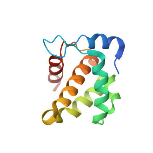
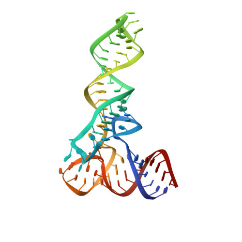
Structure of HIV-1 matrix domain bound to human tRNALys3


Experimental Data Snapshot

  • Method: X-RAY DIFFRACTION
  • Resolution: 3.15 Å
  • R-Value Free: 0.223 
  • R-Value Work: 0.185 
  • R-Value Observed: 0.189 

Starting Models: experimental
View more details

wwPDB Validation   3D Report Full Report


This is version 1.5 of the entry. See complete history


Literature

HIV-1 matrix-tRNA complex structure reveals basis for host control of Gag localization.

Bou-Nader, C.Muecksch, F.Brown, J.B.Gordon, J.M.York, A.Peng, C.Ghirlando, R.Summers, M.F.Bieniasz, P.D.Zhang, J.

(2021) Cell Host Microbe 29: 1421

  • DOI: https://doi.org/10.1016/j.chom.2021.07.006
  • Primary Citation of Related Structures:  
    7MRL

  • PubMed Abstract: 

    The HIV-1 virion structural polyprotein, Gag, is directed to particle assembly sites at the plasma membrane by its N-terminal matrix (MA) domain. MA also binds to host tRNAs. To understand the molecular basis of MA-tRNA interaction and its potential function, we present a co-crystal structure of HIV-1 MA-tRNA Lys3 complex. The structure reveals a specialized group of MA basic and aromatic residues preconfigured to recognize the distinctive structure of the tRNA elbow. Mutational, cross-linking, fluorescence, and NMR analyses show that the crystallographically defined interface drives MA-tRNA binding in solution and living cells. The structure indicates that MA is unlikely to bind tRNA and membrane simultaneously. Accordingly, single-amino-acid substitutions that abolish MA-tRNA binding caused striking redistribution of Gag to the plasma membrane and reduced HIV-1 replication. Thus, HIV-1 exploits host tRNAs to occlude a membrane localization signal and control the subcellular distribution of its major structural protein.


  • Organizational Affiliation

    Laboratory of Molecular Biology, National Institute of Diabetes and Digestive and Kidney Diseases, Bethesda, MD 20892, USA.


Macromolecules

Find similar proteins by:  (by identity cutoff)  |  3D Structure
Entity ID: 1
MoleculeChains Sequence LengthOrganismDetailsImage
HIV-1 matrix domainA [auth C],
C [auth B]
108Human immunodeficiency virus 1Mutation(s): 0 
Gene Names: gag
UniProt
Find proteins for C1JB69 (Human immunodeficiency virus 1)
Explore C1JB69 
Go to UniProtKB:  C1JB69
Entity Groups  
Sequence Clusters30% Identity50% Identity70% Identity90% Identity95% Identity100% Identity
UniProt GroupC1JB69
Sequence Annotations
Expand
  • Reference Sequence
Find similar nucleic acids by:  (by identity cutoff)  |  3D Structure
Entity ID: 2
MoleculeChains LengthOrganismImage
tRNA Lys3B [auth A]75Homo sapiens
Sequence Annotations
Expand
  • Reference Sequence
Small Molecules
Ligands 1 Unique
IDChains Name / Formula / InChI Key2D Diagram3D Interactions
MG
Query on MG

Download Ideal Coordinates CCD File 
D [auth A]MAGNESIUM ION
Mg
JLVVSXFLKOJNIY-UHFFFAOYSA-N
Experimental Data & Validation

Experimental Data

  • Method: X-RAY DIFFRACTION
  • Resolution: 3.15 Å
  • R-Value Free: 0.223 
  • R-Value Work: 0.185 
  • R-Value Observed: 0.189 
  • Space Group: I 2 2 2
Unit Cell:
Length ( Å )Angle ( ˚ )
a = 96.717α = 90
b = 108.846β = 90
c = 146.021γ = 90
Software Package:
Software NamePurpose
PHENIXrefinement
MOSFLMdata reduction
xia2data scaling
PHASERphasing
PDB_EXTRACTdata extraction

Structure Validation

View Full Validation Report


Entry History & Funding Information

Deposition Data


Funding OrganizationLocationGrant Number
National Institutes of Health/National Institute of Diabetes and Digestive and Kidney Disease (NIH/NIDDK)United States--

Revision History  (Full details and data files)

  • Version 1.0: 2021-08-11
    Type: Initial release
  • Version 1.1: 2021-08-18
    Changes: Derived calculations
  • Version 1.2: 2021-08-25
    Changes: Database references
  • Version 1.3: 2021-09-22
    Changes: Database references
  • Version 1.4: 2023-06-07
    Changes: Derived calculations, Refinement description
  • Version 1.5: 2023-10-25
    Changes: Data collection, Refinement description