7MLU

Cryo-EM reveals partially and fully assembled native glycine receptors,homomeric pentamer


Experimental Data Snapshot

  • Method: ELECTRON MICROSCOPY
  • Resolution: 4.10 Å
  • Aggregation State: PARTICLE 
  • Reconstruction Method: SINGLE PARTICLE 

wwPDB Validation   3D Report Full Report


This is version 1.2 of the entry. See complete history


Literature

Architecture and assembly mechanism of native glycine receptors.

Zhu, H.Gouaux, E.

(2021) Nature 599: 513-517

  • DOI: https://doi.org/10.1038/s41586-021-04022-z
  • Primary Citation of Related Structures:  
    7MLU, 7MLV, 7MLY

  • PubMed Abstract: 

    Glycine receptors (GlyRs) are pentameric, 'Cys-loop' receptors that form chloride-permeable channels and mediate fast inhibitory signalling throughout the central nervous system 1,2 . In the spinal cord and brainstem, GlyRs regulate locomotion and cause movement disorders when mutated 2,3 . However, the stoichiometry of native GlyRs and the mechanism by which they are assembled remain unclear, despite extensive investigation 4-8 . Here we report cryo-electron microscopy structures of native GlyRs from pig spinal cord and brainstem, revealing structural insights into heteromeric receptors and their predominant subunit stoichiometry of 4α:1β. Within the heteromeric pentamer, the β(+)-α(-) interface adopts a structure that is distinct from the α(+)-α(-) and α(+)-β(-) interfaces. Furthermore, the β-subunit contains a unique phenylalanine residue that resides within the pore and disrupts the canonical picrotoxin site. These results explain why inclusion of the β-subunit breaks receptor symmetry and alters ion channel pharmacology. We also find incomplete receptor complexes and, by elucidating their structures, reveal the architectures of partially assembled α-trimers and α-tetramers.


  • Organizational Affiliation

    Vollum Institute, Oregon Health and Science University, Portland, OR, USA.


Macromolecules
Find similar proteins by:  (by identity cutoff)  |  3D Structure
Entity ID: 1
MoleculeChains Sequence LengthOrganismDetailsImage
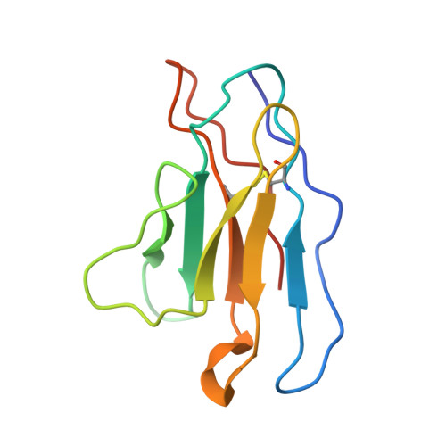
3D1 Fab Light ChainA [auth K],
C [auth J],
E [auth O],
G [auth M],
I
107Rattus norvegicusMutation(s): 0 
Entity Groups  
Sequence Clusters30% Identity50% Identity70% Identity90% Identity95% Identity100% Identity
Sequence Annotations
Expand
  • Reference Sequence
Find similar proteins by:  (by identity cutoff)  |  3D Structure
Entity ID: 2
MoleculeChains Sequence LengthOrganismDetailsImage
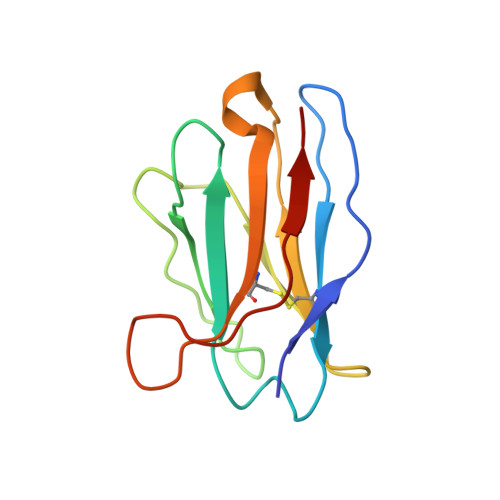
3D1 Fab Heavy ChainB [auth F],
D [auth G],
F [auth N],
H [auth L],
J [auth H]
118Rattus norvegicusMutation(s): 0 
Entity Groups  
Sequence Clusters30% Identity50% Identity70% Identity90% Identity95% Identity100% Identity
Sequence Annotations
Expand
  • Reference Sequence
Find similar proteins by:  (by identity cutoff)  |  3D Structure
Entity ID: 3
MoleculeChains Sequence LengthOrganismDetailsImage
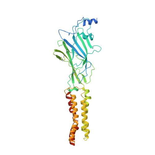
Glycine receptor alpha 1K [auth A],
L [auth E],
M [auth D],
N [auth B],
O [auth C]
456Sus scrofaMutation(s): 0 
Membrane Entity: Yes 
UniProt
Find proteins for A0A4X1TUM3 (Sus scrofa)
Explore A0A4X1TUM3 
Go to UniProtKB:  A0A4X1TUM3
Entity Groups  
Sequence Clusters30% Identity50% Identity70% Identity90% Identity95% Identity100% Identity
UniProt GroupA0A4X1TUM3
Sequence Annotations
Expand
  • Reference Sequence
Experimental Data & Validation

Experimental Data

  • Method: ELECTRON MICROSCOPY
  • Resolution: 4.10 Å
  • Aggregation State: PARTICLE 
  • Reconstruction Method: SINGLE PARTICLE 
EM Software:
TaskSoftware PackageVersion
RECONSTRUCTIONcryoSPARC

Structure Validation

View Full Validation Report


Entry History & Funding Information

Deposition Data


Funding OrganizationLocationGrant Number
National Institutes of Health/National Institute of General Medical Sciences (NIH/NIGMS)United StatesR01 GM100400

Revision History  (Full details and data files)

  • Version 1.0: 2021-09-29
    Type: Initial release
  • Version 1.1: 2021-10-06
    Changes: Database references
  • Version 1.2: 2021-12-01
    Changes: Database references