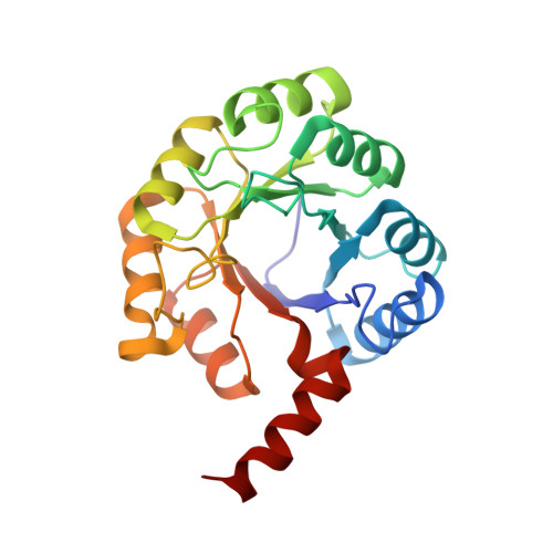
N-acetylmannosamine-6-phosphate 2-epimerase uses a novel substrate-assisted mechanism to catalyze amino sugar epimerization.
Currie, M.J., Manjunath, L., Horne, C.R., Rendle, P.M., Subramanian, R., Friemann, R., Fairbanks, A.J., Muscroft-Taylor, A.C., North, R.A., Dobson, R.C.J.(2021) J Biol Chem 297: 101113-101113
- PubMed: 34437902 Search on PubMedSearch on PubMed Central
- DOI: https://doi.org/10.1016/j.jbc.2021.101113
- Primary Citation Related Structures:
6VVA, 7MFN, 7MFS, 7MQT - PubMed Abstract:
There are five known general catalytic mechanisms used by enzymes to catalyze carbohydrate epimerization. The amino sugar epimerase N-acetylmannosamine-6-phosphate 2-epimerase (NanE) has been proposed to use a deprotonation-reprotonation mechanism, with an essential catalytic lysine required for both steps. However, the structural determinants of this mechanism are not clearly established. We characterized NanE from Staphylococcus aureus using a new coupled assay to monitor NanE catalysis in real time and found that it has kinetic constants comparable with other species. The crystal structure of NanE from Staphylococcus aureus, which comprises a triosephosphate isomerase barrel fold with an unusual dimeric architecture, was solved with both natural and modified substrates. Using these substrate-bound structures, we identified the following active-site residues lining the cleft at the C-terminal end of the β-strands: Gln11, Arg40, Lys63, Asp124, Glu180, and Arg208, which were individually substituted and assessed in relation to the mechanism. From this, we re-evaluated the central role of Glu180 in this mechanism alongside the catalytic lysine. We observed that the substrate is bound in a conformation that ideally positions the C5 hydroxyl group to be activated by Glu180 and donate a proton to the C2 carbon. Taken together, we propose that NanE uses a novel substrate-assisted proton displacement mechanism to invert the C2 stereocenter of N-acetylmannosamine-6-phosphate. Our data and mechanistic interpretation may be useful in the development of inhibitors of this enzyme or in enzyme engineering to produce biocatalysts capable of changing the stereochemistry of molecules that are not amenable to synthetic methods.
- Biomolecular Interaction Centre and School of Biological Sciences, University of Canterbury, Christchurch, New Zealand.
Organizational Affiliation: