7MEQ

Crystal structure of human TMPRSS2 in complex with Nafamostat


Experimental Data Snapshot

  • Method: X-RAY DIFFRACTION
  • Resolution: 1.95 Å
  • R-Value Free: 0.225 
  • R-Value Work: 0.192 
  • R-Value Observed: 0.193 

wwPDB Validation   3D Report Full Report


Ligand Structure Quality Assessment 


This is version 1.4 of the entry. See complete history


Literature

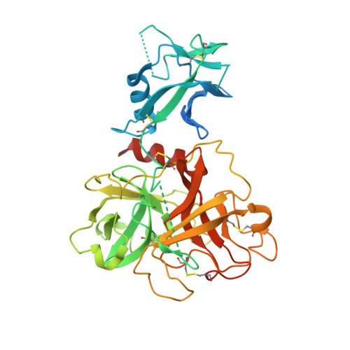
Structure and activity of human TMPRSS2 protease implicated in SARS-CoV-2 activation.

Fraser, B.J.Beldar, S.Seitova, A.Hutchinson, A.Mannar, D.Li, Y.Kwon, D.Tan, R.Wilson, R.P.Leopold, K.Subramaniam, S.Halabelian, L.Arrowsmith, C.H.Benard, F.

(2022) Nat Chem Biol 18: 963-971

  • DOI: https://doi.org/10.1038/s41589-022-01059-7
  • Primary Citation of Related Structures:  
    7MEQ

  • PubMed Abstract: 

    Transmembrane protease, serine 2 (TMPRSS2) has been identified as key host cell factor for viral entry and pathogenesis of SARS-CoV-2. Specifically, TMPRSS2 proteolytically processes the SARS-CoV-2 Spike (S) protein, enabling virus-host membrane fusion and infection of the airways. We present here a recombinant production strategy for enzymatically active TMPRSS2 and characterization of its matured proteolytic activity, as well as its 1.95 Å X-ray cocrystal structure with the synthetic protease inhibitor nafamostat. Our study provides a structural basis for the potent but nonspecific inhibition by nafamostat and identifies distinguishing features of the TMPRSS2 substrate binding pocket that explain specificity. TMPRSS2 cleaved SARS-CoV-2 S protein at multiple sites, including the canonical S1/S2 cleavage site. We ranked the potency of clinical protease inhibitors with half-maximal inhibitory concentrations ranging from 1.4 nM to 120 µM and determined inhibitor mechanisms of action, providing the groundwork for drug development efforts to selectively inhibit TMPRSS2.


  • Organizational Affiliation

    Department of Molecular Oncology, British Columbia Cancer Research Institute, Vancouver, British Columbia, Canada.


Macromolecules
Find similar proteins by:  (by identity cutoff)  |  3D Structure
Entity ID: 1
MoleculeChains Sequence LengthOrganismDetailsImage
Transmembrane protease serine 2395Homo sapiensMutation(s): 0 
Gene Names: TMPRSS2PRSS10
EC: 3.4.21
UniProt & NIH Common Fund Data Resources
Find proteins for O15393 (Homo sapiens)
Explore O15393 
Go to UniProtKB:  O15393
PHAROS:  O15393
GTEx:  ENSG00000184012 
Entity Groups  
Sequence Clusters30% Identity50% Identity70% Identity90% Identity95% Identity100% Identity
UniProt GroupO15393
Sequence Annotations
Expand
  • Reference Sequence
Oligosaccharides

Help

Entity ID: 2
MoleculeChains Length2D Diagram Glycosylation3D Interactions
2-acetamido-2-deoxy-beta-D-glucopyranose-(1-4)-2-acetamido-2-deoxy-beta-D-glucopyranose
B
2N-Glycosylation
Glycosylation Resources
GlyTouCan:  G42666HT
GlyCosmos:  G42666HT
GlyGen:  G42666HT
Experimental Data & Validation

Experimental Data

  • Method: X-RAY DIFFRACTION
  • Resolution: 1.95 Å
  • R-Value Free: 0.225 
  • R-Value Work: 0.192 
  • R-Value Observed: 0.193 
  • Space Group: P 1 21 1
Unit Cell:
Length ( Å )Angle ( ˚ )
a = 59.445α = 90
b = 51.425β = 91.53
c = 64.353γ = 90
Software Package:
Software NamePurpose
BUSTERrefinement
XDSdata reduction
Aimlessdata scaling
PHASERphasing
PDB_EXTRACTdata extraction

Structure Validation

View Full Validation Report


Ligand Structure Quality Assessment 


Entry History 

Revision History  (Full details and data files)

  • Version 1.0: 2021-04-21
    Type: Initial release
  • Version 1.1: 2021-11-03
    Changes: Database references
  • Version 1.2: 2022-06-22
    Changes: Database references
  • Version 1.3: 2022-09-07
    Changes: Database references
  • Version 1.4: 2023-10-18
    Changes: Data collection, Database references, Refinement description