7MCH

Crystal structure of a single-chain E/F type bilin lyase-isomerase MpeQ in space group C2221


Experimental Data Snapshot

  • Method: X-RAY DIFFRACTION
  • Resolution: 2.95 Å
  • R-Value Free: 0.308 
  • R-Value Work: 0.244 
  • R-Value Observed: 0.249 

wwPDB Validation   3D Report Full Report


This is version 1.0 of the entry. See complete history


Literature

Crystal structure and molecular mechanism of an E/F type bilin lyase-isomerase.

Kumarapperuma, I.Joseph, K.L.Wang, C.Biju, L.M.Tom, I.P.Weaver, K.D.Grebert, T.Partensky, F.Schluchter, W.M.Yang, X.

(2022) Structure 30: 564-574.e3

  • DOI: https://doi.org/10.1016/j.str.2022.01.007
  • Primary Citation of Related Structures:  
    7MC4, 7MCH

  • PubMed Abstract: 

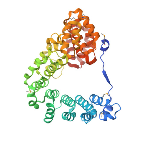
    Chromophore attachment of the light-harvesting apparatus represents one of the most important post-translational modifications in photosynthetic cyanobacteria. Extensive pigment diversity of cyanobacteria critically depends on bilin lyases that covalently attach chemically distinct chromophores to phycobiliproteins. However, how bilin lyases catalyze bilin ligation reactions and how some lyases acquire additional isomerase abilities remain elusive at the molecular level. Here, we report the crystal structure of a representative bilin lyase-isomerase MpeQ. This structure has revealed a "question-mark" protein architecture that unambiguously establishes the active site conserved among the E/F-type bilin lyases. Based on structural, mutational, and modeling data, we demonstrate that stereoselectivity of the active site plays a critical role in conferring the isomerase activity of MpeQ. We further advance a tyrosine-mediated reaction scheme unifying different types of bilin lyases. These results suggest that lyases and isomerase actions of bilin lyases arise from two coupled molecular events of distinct origin.


  • Organizational Affiliation

    Department of Chemistry, University of Illinois Chicago, Chicago, IL 60607, USA.


Macromolecules
Find similar proteins by:  (by identity cutoff)  |  3D Structure
Entity ID: 1
MoleculeChains Sequence LengthOrganismDetailsImage
bilin lyase-isomerase
A, B
416Synechococcus sp. A15-62Mutation(s): 0 
Gene Names: mpeY
UniProt
Find proteins for U3MW57 (Synechococcus sp. A15-62)
Explore U3MW57 
Go to UniProtKB:  U3MW57
Entity Groups  
Sequence Clusters30% Identity50% Identity70% Identity90% Identity95% Identity100% Identity
UniProt GroupU3MW57
Sequence Annotations
Expand
  • Reference Sequence
Small Molecules
Modified Residues  1 Unique
IDChains TypeFormula2D DiagramParent
MSE
Query on MSE
A, B
L-PEPTIDE LINKINGC5 H11 N O2 SeMET
Experimental Data & Validation

Experimental Data

  • Method: X-RAY DIFFRACTION
  • Resolution: 2.95 Å
  • R-Value Free: 0.308 
  • R-Value Work: 0.244 
  • R-Value Observed: 0.249 
  • Space Group: C 2 2 21
Unit Cell:
Length ( Å )Angle ( ˚ )
a = 136.875α = 90
b = 174.3β = 90
c = 113.857γ = 90
Software Package:
Software NamePurpose
PHENIXrefinement
PDB_EXTRACTdata extraction
HKL-2000data reduction
HKL-2000data scaling
AutoSolphasing

Structure Validation

View Full Validation Report


Entry History & Funding Information

Deposition Data


Funding OrganizationLocationGrant Number
National Institutes of Health/National Eye Institute (NIH/NEI)United StatesEY024363

Revision History  (Full details and data files)

  • Version 1.0: 2022-08-24
    Type: Initial release