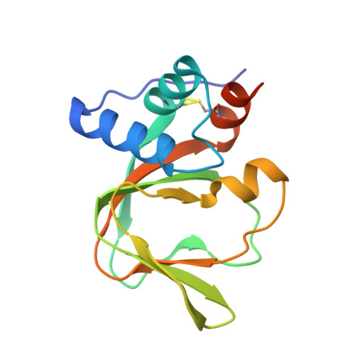
An auto-inhibited state of protein kinase G and implications for selective activation.
Sharma, R., Kim, J.J., Qin, L., Henning, P., Akimoto, M., VanSchouwen, B., Kaur, G., Sankaran, B., MacKenzie, K.R., Melacini, G., Casteel, D.E., Herberg, F.W., Kim, C.W.(2022) Elife 11
- PubMed: 35929723
- DOI: https://doi.org/10.7554/eLife.79530
- Primary Citation of Related Structures:
7LV3, 7MBJ - PubMed Abstract:
Cyclic GMP-dependent protein kinases (PKGs) are key mediators of the nitric oxide/cyclic guanosine monophosphate (cGMP) signaling pathway that regulates biological functions as diverse as smooth muscle contraction, cardiac function, and axon guidance. Understanding how cGMP differentially triggers mammalian PKG isoforms could lead to new therapeutics that inhibit or activate PKGs, complementing drugs that target nitric oxide synthases and cyclic nucleotide phosphodiesterases in this signaling axis. Alternate splicing of PRKG1 transcripts confers distinct leucine zippers, linkers, and auto-inhibitory (AI) pseudo-substrate sequences to PKG Iα and Iβ that result in isoform-specific activation properties, but the mechanism of enzyme auto-inhibition and its alleviation by cGMP is not well understood. Here, we present a crystal structure of PKG Iβ in which the AI sequence and the cyclic nucleotide-binding (CNB) domains are bound to the catalytic domain, providing a snapshot of the auto-inhibited state. Specific contacts between the PKG Iβ AI sequence and the enzyme active site help explain isoform-specific activation constants and the effects of phosphorylation in the linker. We also present a crystal structure of a PKG I CNB domain with an activating mutation linked to Thoracic Aortic Aneurysms and Dissections. Similarity of this structure to wildtype cGMP-bound domains and differences with the auto-inhibited enzyme provide a mechanistic basis for constitutive activation. We show that PKG Iβ auto-inhibition is mediated by contacts within each monomer of the native full-length dimeric protein, and using the available structural and biochemical data we develop a model for the regulation and cooperative activation of PKGs.
- Department of Pharmacology and Chemical Biology and Center for Drug Discovery, Baylor College of Medicine, Houston, United States.
Organizational Affiliation:


