7M8W | pdb_00007m8w

XFEL crystal structure of the prostaglandin D2 receptor CRTH2 in complex with 15R-methyl-PGD2


Experimental Data Snapshot

  • Method: X-RAY DIFFRACTION
  • Resolution: 2.61 Å
  • R-Value Free: 
    0.267 (Depositor), 0.264 (DCC) 
  • R-Value Work: 
    0.229 (Depositor), 0.231 (DCC) 
  • R-Value Observed: 
    0.231 (Depositor) 

Starting Model: experimental
View more details

wwPDB Validation 3D Report Full Report

Validation slider image for 7M8W

Ligand Structure Quality Assessment 


This is version 1.1 of the entry. See complete history

Literature

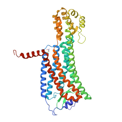
Molecular basis for lipid recognition by the prostaglandin D 2 receptor CRTH2.

Liu, H.Deepak, R.N.V.K.Shiriaeva, A.Gati, C.Batyuk, A.Hu, H.Weierstall, U.Liu, W.Wang, L.Cherezov, V.Fan, H.Zhang, C.

(2021) Proc Natl Acad Sci U S A 118

  • DOI: https://doi.org/10.1073/pnas.2102813118
  • Primary Citation Related Structures: 
    7M8W

  • PubMed Abstract: 

    Prostaglandin D 2 (PGD 2 ) signals through the G protein-coupled receptor (GPCR) CRTH2 to mediate various inflammatory responses. CRTH2 is the only member of the prostanoid receptor family that is phylogenetically distant from others, implying a nonconserved mechanism of lipid action on CRTH2. Here, we report a crystal structure of human CRTH2 bound to a PGD 2 derivative, 15R-methyl-PGD 2 (15mPGD 2 ), by serial femtosecond crystallography. The structure revealed a "polar group in"-binding mode of 15mPGD 2 contrasting the "polar group out"-binding mode of PGE 2 in its receptor EP3. Structural comparison analysis suggested that these two lipid-binding modes, associated with distinct charge distributions of ligand-binding pockets, may apply to other lipid GPCRs. Molecular dynamics simulations together with mutagenesis studies also identified charged residues at the ligand entry port that function to capture lipid ligands of CRTH2 from the lipid bilayer. Together, our studies suggest critical roles of charge environment in lipid recognition by GPCRs.


  • Organizational Affiliation
    • Department of Pharmacology and Chemical Biology, School of Medicine, University of Pittsburgh, Pittsburgh, PA 15261.

Macromolecule Content 

  • Total Structure Weight: 53.29 kDa 
  • Atom Count: 3,501 
  • Modeled Residue Count: 437 
  • Deposited Residue Count: 470 
  • Unique protein chains: 1

Macromolecules

Find similar proteins by:|  3D Structure
Entity ID: 1
MoleculeChains  Sequence LengthOrganismDetailsImage
Prostaglandin D2 receptor 2, Endolysin chimera470Homo sapiensTequatrovirus T4Mutation(s): 3 
Gene Names: PTGDR2CRTH2DL1RGPR44eT4Tp126
EC: 3.2.1.17
Membrane Entity: Yes 
UniProt & NIH Common Fund Data Resources
Find proteins for D9IEF7 (Enterobacteria phage T4)
Explore D9IEF7 
Go to UniProtKB:  D9IEF7
Find proteins for Q9Y5Y4 (Homo sapiens)
Explore Q9Y5Y4 
Go to UniProtKB:  Q9Y5Y4
PHAROS:  Q9Y5Y4
GTEx:  ENSG00000183134 
Entity Groups
Sequence Clusters30% Identity50% Identity70% Identity90% Identity95% Identity100% Identity
UniProt GroupsQ9Y5Y4D9IEF7
Sequence Annotations
Expand
Reference Sequence

Small Molecules

Ligands 4 Unique
IDChains Name / Formula / InChI Key2D Diagram3D Interactions
YSS
(Subject of Investigation/LOI)

Query on YSS


Download:Ideal Coordinates CCD File
B [auth A]15R-methyl-prostaglandin D2
C21 H34 O5
CTXLUMAOXBULOZ-BKVRKCTKSA-N
FLC

Query on FLC


Download:Ideal Coordinates CCD File
D [auth A]CITRATE ANION
C6 H5 O7
KRKNYBCHXYNGOX-UHFFFAOYSA-K
SO4

Query on SO4


Download:Ideal Coordinates CCD File
E [auth A],
F [auth A],
G [auth A],
H [auth A],
I [auth A]
SULFATE ION
O4 S
QAOWNCQODCNURD-UHFFFAOYSA-L
NA

Query on NA


Download:Ideal Coordinates CCD File
C [auth A]SODIUM ION
Na
FKNQFGJONOIPTF-UHFFFAOYSA-N
Modified Residues  1 Unique
IDChains TypeFormula2D DiagramParent
YCM
Query on YCM
A
L-PEPTIDE LINKINGC5 H10 N2 O3 SCYS

Experimental Data & Validation

Experimental Data

  • Method: X-RAY DIFFRACTION
  • Resolution: 2.61 Å
  • R-Value Free:  0.267 (Depositor), 0.264 (DCC) 
  • R-Value Work:  0.229 (Depositor), 0.231 (DCC) 
  • R-Value Observed: 0.231 (Depositor) 
Space Group: P 21 21 21
Unit Cell:
Length ( Å )Angle ( ˚ )
a = 51.4α = 90
b = 67.8β = 90
c = 265.4γ = 90
Software Package:
Software NamePurpose
PHENIXrefinement
PDB_EXTRACTdata extraction
CrystFELdata reduction
CrystFELdata scaling
PHASERphasing

Structure Validation

View Full Validation Report


Ligand Structure Quality Assessment 


Entry History 

Deposition Data

Revision History  (Full details and data files)

  • Version 1.0: 2021-08-25
    Type: Initial release
  • Version 1.1: 2023-10-18
    Changes: Data collection, Refinement description