7M7J | pdb_00007m7j

6-Deoxyerythronolide B synthase (DEBS) module 1 in complex with antibody fragment 1B2: "turnstile closed" state (TE-free)


Experimental Data Snapshot

  • Method: ELECTRON MICROSCOPY
  • Resolution: 4.30 Å
  • Aggregation State: PARTICLE 
  • Reconstruction Method: SINGLE PARTICLE 

wwPDB Validation 3D Report Full Report

Validation slider image for 7M7J

This is version 1.2 of the entry. See complete history

Literature

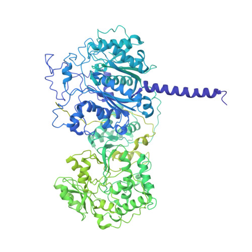
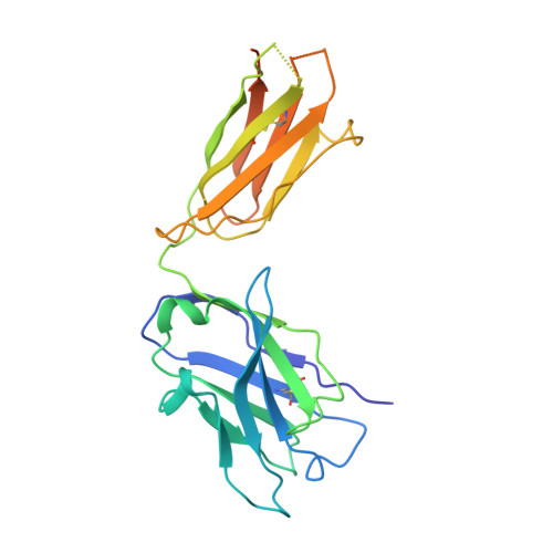
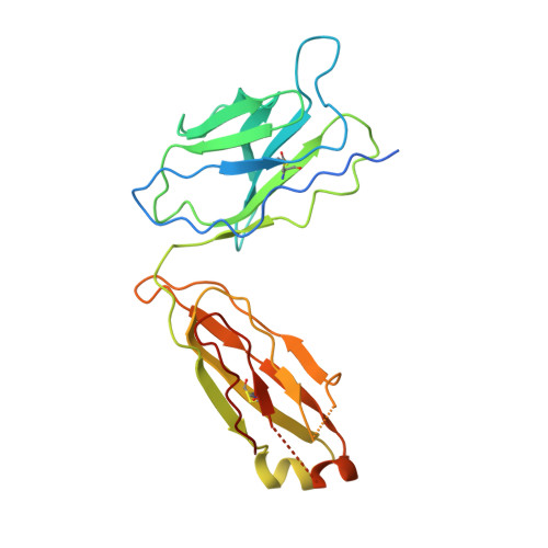
Mapping the catalytic conformations of an assembly-line polyketide synthase module.

Cogan, D.P.Zhang, K.Li, X.Li, S.Pintilie, G.D.Roh, S.H.Craik, C.S.Chiu, W.Khosla, C.

(2021) Science 374: 729-734

  • DOI: https://doi.org/10.1126/science.abi8358
  • Primary Citation Related Structures: 
    7M7E, 7M7F, 7M7G, 7M7H, 7M7I, 7M7J

  • PubMed Abstract: 

    Assembly-line polyketide synthases, such as the 6-deoxyerythronolide B synthase (DEBS), are large enzyme factories prized for their ability to produce specific and complex polyketide products. By channeling protein-tethered substrates across multiple active sites in a defined linear sequence, these enzymes facilitate programmed small-molecule syntheses that could theoretically be harnessed to access countless polyketide product structures. Using cryogenic electron microscopy to study DEBS module 1, we present a structural model describing this substrate-channeling phenomenon. Our 3.2- to 4.3-angstrom-resolution structures of the intact module reveal key domain-domain interfaces and highlight an unexpected module asymmetry. We also present the structure of a product-bound module that shines light on a recently described “turnstile” mechanism for transient gating of active sites along the assembly line.


  • Organizational Affiliation
    • Department of Chemistry, Stanford University, Stanford, CA 94305, USA.

Macromolecule Content 

  • Total Structure Weight: 439.8 kDa 
  • Atom Count: 23,224 
  • Modeled Residue Count: 3,124 
  • Deposited Residue Count: 4,156 
  • Unique protein chains: 3

Macromolecules

Find similar proteins by:|  3D Structure
Entity ID: 1
MoleculeChains  Sequence LengthOrganismDetailsImage
EryAIA [auth B],
B [auth A]
1,593Saccharopolyspora erythraeaMutation(s): 0 
Gene Names: eryAI
EC: 2.3.1.94
UniProt
Find proteins for Q03131 (Saccharopolyspora erythraea)
Explore Q03131 
Go to UniProtKB:  Q03131
Find proteins for Q5UNP6 (Saccharopolyspora erythraea)
Explore Q5UNP6 
Go to UniProtKB:  Q5UNP6
Entity Groups
Sequence Clusters30% Identity50% Identity70% Identity90% Identity95% Identity100% Identity
UniProt GroupsQ5UNP6Q03131
Sequence Annotations
Expand
Reference Sequence
Find similar proteins by:|  3D Structure
Entity ID: 2
MoleculeChains  Sequence LengthOrganismDetailsImage
1B2 (heavy chain)
C, E
249Homo sapiensMutation(s): 0 
Find similar proteins by:|  3D Structure
Entity ID: 3
MoleculeChains  Sequence LengthOrganismDetailsImage
1B2 (light chain)
D, F
236Homo sapiensMutation(s): 0 

Experimental Data & Validation

Experimental Data

  • Method: ELECTRON MICROSCOPY
  • Resolution: 4.30 Å
  • Aggregation State: PARTICLE 
  • Reconstruction Method: SINGLE PARTICLE 
EM Software:
TaskSoftware PackageVersion
MODEL REFINEMENTPHENIX
RECONSTRUCTIONcryoSPARC

Structure Validation

View Full Validation Report


Entry History 

& Funding Information

Deposition Data


Funding OrganizationLocationGrant Number
National Institutes of Health/National Institute of General Medical Sciences (NIH/NIGMS)United StatesR01GM079429

Revision History  (Full details and data files)

  • Version 1.0: 2021-11-17
    Type: Initial release
  • Version 1.1: 2024-10-16
    Changes: Data collection, Structure summary
  • Version 1.2: 2025-05-21
    Changes: Data collection