


Development, Characterization, and Structural Analysis of a Genetically Encoded Red Fluorescent Peroxynitrite Biosensor
Pang, Y., Huang, M., Zhang, S., Fan, Y., Yeh, H., Xiong, Y., Li, X., Ng, H.L., Ai, H.To be published.
Experimental Data Snapshot
Starting Model: experimental
View more details
wwPDB Validation 3D Report Full Report

Entity ID: 1 | |||||
|---|---|---|---|---|---|
| Molecule | Chains | Sequence Length | Organism | Details | Image |
| Red Fluorescent pnRFP B30Y mutant | 262 | Discosoma sp. | Mutation(s): 0 | 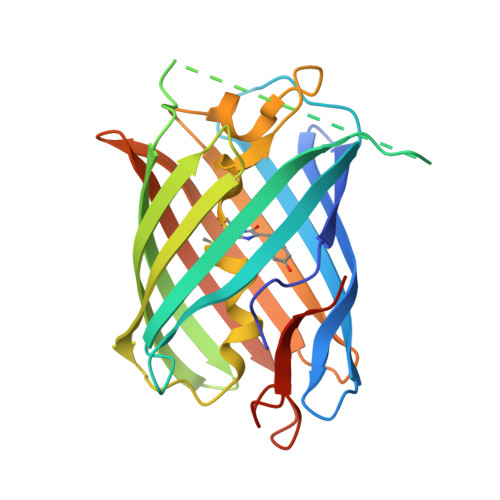 | |
| Ligands 1 Unique | |||||
|---|---|---|---|---|---|
| ID | Chains | Name / Formula / InChI Key | 2D Diagram | 3D Interactions | |
| PO4 Download:Ideal Coordinates CCD File | B [auth A] | PHOSPHATE ION O4 P NBIIXXVUZAFLBC-UHFFFAOYSA-K |  | ||
| Modified Residues 1 Unique | |||||
|---|---|---|---|---|---|
| ID | Chains | Type | Formula | 2D Diagram | Parent |
| NRQ Query on NRQ | A | L-PEPTIDE LINKING | C16 H17 N3 O4 S |  | MET, TYR, GLY |
| Length ( Å ) | Angle ( ˚ ) |
|---|---|
| a = 84.284 | α = 90 |
| b = 34.107 | β = 110.79 |
| c = 89.222 | γ = 90 |
| Software Name | Purpose |
|---|---|
| REFMAC | refinement |
| Aimless | data scaling |
| PDB_EXTRACT | data extraction |
| XDS | data reduction |
| PHASER | phasing |


