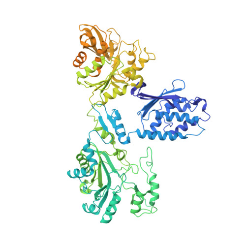
Structures and function of the amino acid polymerase cyanophycin synthetase.
Sharon, I., Haque, A.S., Grogg, M., Lahiri, I., Seebach, D., Leschziner, A.E., Hilvert, D., Schmeing, T.M.(2021) Nat Chem Biol 17: 1101-1110
- PubMed: 34385683
- DOI: https://doi.org/10.1038/s41589-021-00854-y
- Primary Citation of Related Structures:
7LG5, 7LGJ, 7LGM, 7LGN, 7LGQ - PubMed Abstract:
Cyanophycin is a natural biopolymer produced by a wide range of bacteria, consisting of a chain of poly-L-Asp residues with L-Arg residues attached to the β-carboxylate sidechains by isopeptide bonds. Cyanophycin is synthesized from ATP, aspartic acid and arginine by a homooligomeric enzyme called cyanophycin synthetase (CphA1). CphA1 has domains that are homologous to glutathione synthetases and muramyl ligases, but no other structural information has been available. Here, we present cryo-electron microscopy and X-ray crystallography structures of cyanophycin synthetases from three different bacteria, including cocomplex structures of CphA1 with ATP and cyanophycin polymer analogs at 2.6 Å resolution. These structures reveal two distinct tetrameric architectures, show the configuration of active sites and polymer-binding regions, indicate dynamic conformational changes and afford insight into catalytic mechanism. Accompanying biochemical interrogation of substrate binding sites, catalytic centers and oligomerization interfaces combine with the structures to provide a holistic understanding of cyanophycin biosynthesis.
- Department of Biochemistry and Centre de Recherche en Biologie Structurale, McGill University, Montréal, Quebec, Canada.
Organizational Affiliation:


