7LG4 | pdb_00007lg4

Green fluorescent protein from Aequorea macrodactyla - amacGFP


Experimental Data Snapshot

  • Method: X-RAY DIFFRACTION
  • Resolution: 1.81 Å
  • R-Value Free: 
    0.228 (Depositor), 0.239 (DCC) 
  • R-Value Work: 
    0.182 (Depositor), 0.193 (DCC) 
  • R-Value Observed: 
    0.184 (Depositor) 

Starting Model: experimental
View more details

wwPDB Validation 3D Report Full Report

Validation slider image for 7LG4

This is version 3.0 of the entry. See complete history

Literature

Heterogeneity of the GFP fitness landscape and data-driven protein design.

Gonzalez Somermeyer, L.Fleiss, A.Mishin, A.S.Bozhanova, N.G.Igolkina, A.A.Meiler, J.Alaball Pujol, M.E.Putintseva, E.V.Sarkisyan, K.S.Kondrashov, F.A.

(2022) Elife 11

  • DOI: https://doi.org/10.7554/eLife.75842
  • Primary Citation Related Structures: 
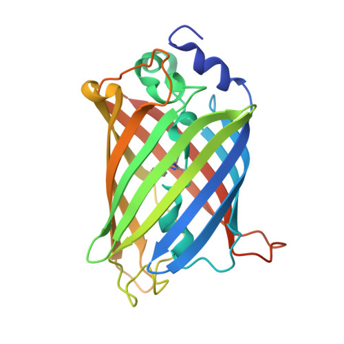
    7LG4

  • PubMed Abstract: 

    Studies of protein fitness landscapes reveal biophysical constraints guiding protein evolution and empower prediction of functional proteins. However, generalisation of these findings is limited due to scarceness of systematic data on fitness landscapes of proteins with a defined evolutionary relationship. We characterized the fitness peaks of four orthologous fluorescent proteins with a broad range of sequence divergence. While two of the four studied fitness peaks were sharp, the other two were considerably flatter, being almost entirely free of epistatic interactions. Mutationally robust proteins, characterized by a flat fitness peak, were not optimal templates for machine-learning-driven protein design - instead, predictions were more accurate for fragile proteins with epistatic landscapes. Our work paves insights for practical application of fitness landscape heterogeneity in protein engineering.


  • Organizational Affiliation
    • Institute of Science and Technology Austria, Klosterneuburg, Austria.

Macromolecule Content 

  • Total Structure Weight: 28.36 kDa 
  • Atom Count: 2,051 
  • Modeled Residue Count: 228 
  • Deposited Residue Count: 242 
  • Unique protein chains: 1

Macromolecules

Find similar proteins by:|  3D Structure
Entity ID: 1
MoleculeChains  Sequence LengthOrganismDetailsImage
Green fluorescent protein242Aequorea macrodactylaMutation(s): 1 
Gene Names: GFP
UniProt
Find proteins for Q8WTC7 (Aequorea macrodactyla)
Explore Q8WTC7 
Go to UniProtKB:  Q8WTC7
Entity Groups
Sequence Clusters30% Identity50% Identity70% Identity90% Identity95% Identity100% Identity
UniProt GroupQ8WTC7
Sequence Annotations
Expand
Reference Sequence

Small Molecules

Ligands 3 Unique
IDChains Name / Formula / InChI Key2D Diagram3D Interactions
PEG

Query on PEG


Download:Ideal Coordinates CCD File
G [auth A]DI(HYDROXYETHYL)ETHER
C4 H10 O3
MTHSVFCYNBDYFN-UHFFFAOYSA-N
GOL

Query on GOL


Download:Ideal Coordinates CCD File
H [auth A]GLYCEROL
C3 H8 O3
PEDCQBHIVMGVHV-UHFFFAOYSA-N
EDO

Query on EDO


Download:Ideal Coordinates CCD File
B [auth A],
C [auth A],
D [auth A],
E [auth A],
F [auth A]
1,2-ETHANEDIOL
C2 H6 O2
LYCAIKOWRPUZTN-UHFFFAOYSA-N
Modified Residues  1 Unique
IDChains TypeFormula2D DiagramParent
GYS
Query on GYS
A
L-PEPTIDE LINKINGC14 H15 N3 O5SER, TYR, GLY

Experimental Data & Validation

Experimental Data

  • Method: X-RAY DIFFRACTION
  • Resolution: 1.81 Å
  • R-Value Free:  0.228 (Depositor), 0.239 (DCC) 
  • R-Value Work:  0.182 (Depositor), 0.193 (DCC) 
  • R-Value Observed: 0.184 (Depositor) 
Space Group: P 1 21 1
Unit Cell:
Length ( Å )Angle ( ˚ )
a = 34.089α = 90
b = 47.488β = 102.01
c = 69.294γ = 90
Software Package:
Software NamePurpose
REFMACrefinement
MOLREPphasing
PROTEUM PLUSdata scaling
PDB_EXTRACTdata extraction
PROTEUM PLUSdata reduction

Structure Validation

View Full Validation Report


Entry History 

Deposition Data

Revision History  (Full details and data files)

  • Version 1.0: 2021-01-27
    Type: Initial release
  • Version 1.1: 2022-05-11
    Changes: Database references
  • Version 1.2: 2022-05-18
    Changes: Database references
  • Version 1.3: 2023-10-18
    Changes: Data collection, Refinement description
  • Version 2.0: 2023-11-15
    Changes: Atomic model, Data collection, Derived calculations
  • Version 2.1: 2024-11-20
    Changes: Structure summary
  • Version 3.0: 2026-03-18
    Changes: Polymer sequence