7L9P

Structure of human SHLD2-SHLD3-REV7-TRIP13(E253Q) complex


Experimental Data Snapshot

  • Method: ELECTRON MICROSCOPY
  • Resolution: 3.60 Å
  • Aggregation State: PARTICLE 
  • Reconstruction Method: SINGLE PARTICLE 

wwPDB Validation   3D Report Full Report


This is version 1.1 of the entry. See complete history


Literature

Molecular mechanisms of assembly and TRIP13-mediated remodeling of the human Shieldin complex.

Xie, W.Wang, S.Wang, J.de la Cruz, M.J.Xu, G.Scaltriti, M.Patel, D.J.

(2021) Proc Natl Acad Sci U S A 118

  • DOI: https://doi.org/10.1073/pnas.2024512118
  • Primary Citation of Related Structures:  
    6WW9, 6WWA, 7L9P

  • PubMed Abstract: 

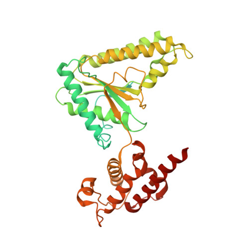
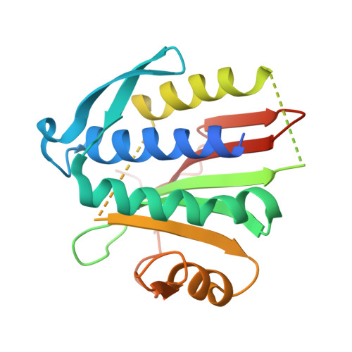
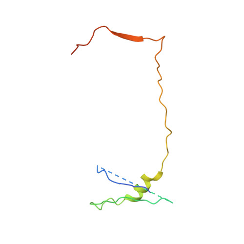
    The Shieldin complex, composed of REV7, SHLD1, SHLD2, and SHLD3, protects DNA double-strand breaks (DSBs) to promote nonhomologous end joining. The AAA + ATPase TRIP13 remodels Shieldin to regulate DNA repair pathway choice. Here we report crystal structures of human SHLD3-REV7 binary and fused SHLD2-SHLD3-REV7 ternary complexes, revealing that assembly of Shieldin requires fused SHLD2-SHLD3 induced conformational heterodimerization of open (O-REV7) and closed (C-REV7) forms of REV7. We also report the cryogenic electron microscopy (cryo-EM) structures of the ATPγS-bound fused SHLD2-SHLD3-REV7-TRIP13 complexes, uncovering the principles underlying the TRIP13-mediated disassembly mechanism of the Shieldin complex. We demonstrate that the N terminus of REV7 inserts into the central channel of TRIP13, setting the stage for pulling the unfolded N-terminal peptide of C-REV7 through the central TRIP13 hexameric channel. The primary interface involves contacts between the safety-belt segment of C-REV7 and a conserved and negatively charged loop of TRIP13. This process is mediated by ATP hydrolysis-triggered rotatory motions of the TRIP13 ATPase, thereby resulting in the disassembly of the Shieldin complex.


  • Organizational Affiliation

    Structural Biology Program, Memorial Sloan Kettering Cancer Center, New York, NY 10065; xiew@mskcc.org pateld@mskcc.org.


Macromolecules
Find similar proteins by:  (by identity cutoff)  |  3D Structure
Entity ID: 1
MoleculeChains Sequence LengthOrganismDetailsImage
Pachytene checkpoint protein 2 homolog
A, B, C, D, E
A, B, C, D, E, F
432Homo sapiensMutation(s): 1 
Gene Names: TRIP13PCH2
UniProt & NIH Common Fund Data Resources
Find proteins for Q15645 (Homo sapiens)
Explore Q15645 
Go to UniProtKB:  Q15645
PHAROS:  Q15645
GTEx:  ENSG00000071539 
Entity Groups  
Sequence Clusters30% Identity50% Identity70% Identity90% Identity95% Identity100% Identity
UniProt GroupQ15645
Sequence Annotations
Expand
  • Reference Sequence
Find similar proteins by:  (by identity cutoff)  |  3D Structure
Entity ID: 2
MoleculeChains Sequence LengthOrganismDetailsImage
Mitotic spindle assembly checkpoint protein MAD2BG,
H [auth I],
I [auth J],
J [auth K]
211Homo sapiensMutation(s): 0 
Gene Names: MAD2L2MAD2BREV7
UniProt & NIH Common Fund Data Resources
Find proteins for Q9UI95 (Homo sapiens)
Explore Q9UI95 
Go to UniProtKB:  Q9UI95
PHAROS:  Q9UI95
GTEx:  ENSG00000116670 
Entity Groups  
Sequence Clusters30% Identity50% Identity70% Identity90% Identity95% Identity100% Identity
UniProt GroupQ9UI95
Sequence Annotations
Expand
  • Reference Sequence
Find similar proteins by:  (by identity cutoff)  |  3D Structure
Entity ID: 3
MoleculeChains Sequence LengthOrganismDetailsImage
Shieldin complex subunit 2, Shieldin complex subunit 3 chimeraK [auth Y],
L [auth X]
99Homo sapiensMutation(s): 0 
Gene Names: SHLD2FAM35ARINN2SHLD3FLJ26957RINN1
UniProt & NIH Common Fund Data Resources
Find proteins for Q86V20 (Homo sapiens)
Explore Q86V20 
Go to UniProtKB:  Q86V20
PHAROS:  Q86V20
GTEx:  ENSG00000122376 
Find proteins for Q6ZNX1 (Homo sapiens)
Explore Q6ZNX1 
Go to UniProtKB:  Q6ZNX1
PHAROS:  Q6ZNX1
GTEx:  ENSG00000253251 
Entity Groups  
Sequence Clusters30% Identity50% Identity70% Identity90% Identity95% Identity100% Identity
UniProt GroupsQ86V20Q6ZNX1
Sequence Annotations
Expand
  • Reference Sequence
Experimental Data & Validation

Experimental Data

  • Method: ELECTRON MICROSCOPY
  • Resolution: 3.60 Å
  • Aggregation State: PARTICLE 
  • Reconstruction Method: SINGLE PARTICLE 

Structure Validation

View Full Validation Report


Entry History 

Deposition Data

Revision History  (Full details and data files)

  • Version 1.0: 2021-03-03
    Type: Initial release
  • Version 1.1: 2024-03-06
    Changes: Data collection, Database references