7L8H

EV68 3C protease (3Cpro) in Complex with Rupintrivir


Experimental Data Snapshot

  • Method: X-RAY DIFFRACTION
  • Resolution: 1.95 Å
  • R-Value Free: 0.186 
  • R-Value Work: 0.158 
  • R-Value Observed: 0.181 

wwPDB Validation   3D Report Full Report


Ligand Structure Quality Assessment 


This is version 1.2 of the entry. See complete history


Literature

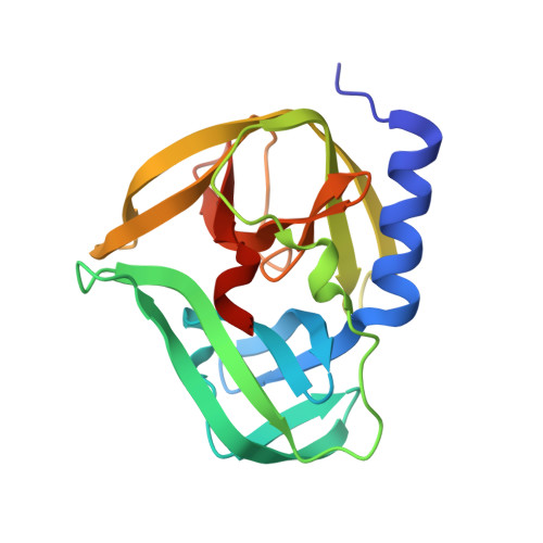
Pan-3C Protease Inhibitor Rupintrivir Binds SARS-CoV-2 Main Protease in a Unique Binding Mode.

Lockbaum, G.J.Henes, M.Lee, J.M.Timm, J.Nalivaika, E.A.Thompson, P.R.Kurt Yilmaz, N.Schiffer, C.A.

(2021) Biochemistry 60: 2925-2931

  • DOI: https://doi.org/10.1021/acs.biochem.1c00414
  • Primary Citation of Related Structures:  
    7L8H, 7L8I, 7L8J

  • PubMed Abstract: 

    Rupintrivir targets the 3C cysteine proteases of the picornaviridae family, which includes rhinoviruses and enteroviruses that cause a range of human diseases. Despite being a pan-3C protease inhibitor, rupintrivir activity is extremely weak against the homologous 3C-like protease of SARS-CoV-2. In this study, the crystal structures of rupintrivir were determined bound to enterovirus 68 (EV68) 3C protease and the 3C-like main protease (M pro ) from SARS-CoV-2. While the EV68 3C protease-rupintrivir structure was similar to previously determined complexes with other picornavirus 3C proteases, rupintrivir bound in a unique conformation to the active site of SARS-CoV-2 M pro splitting the catalytic cysteine and histidine residues. This bifurcation of the catalytic dyad may provide a novel approach for inhibiting cysteine proteases.


  • Organizational Affiliation

    Department of Biochemistry and Molecular Pharmacology, University of Massachusetts Medical School, Worcester, Massachusetts 01605, United States.


Macromolecules
Find similar proteins by:  (by identity cutoff)  |  3D Structure
Entity ID: 1
MoleculeChains Sequence LengthOrganismDetailsImage
3C Protease
A, B
189enterovirus D68Mutation(s): 0 
UniProt
Find proteins for A0A097BW12 (Human enterovirus D68)
Explore A0A097BW12 
Go to UniProtKB:  A0A097BW12
Entity Groups  
Sequence Clusters30% Identity50% Identity70% Identity90% Identity95% Identity100% Identity
UniProt GroupA0A097BW12
Sequence Annotations
Expand
  • Reference Sequence
Small Molecules
Ligands 1 Unique
IDChains Name / Formula / InChI Key2D Diagram3D Interactions
AG7 (Subject of Investigation/LOI)
Query on AG7

Download Ideal Coordinates CCD File 
C [auth A],
D [auth B]
4-{2-(4-FLUORO-BENZYL)-6-METHYL-5-[(5-METHYL-ISOXAZOLE-3-CARBONYL)-AMINO]-4-OXO-HEPTANOYLAMINO}-5-(2-OXO-PYRROLIDIN-3-YL)-PENTANOIC ACID ETHYL ESTER
C31 H41 F N4 O7
LMIUALQNZXJHOG-IFILWLFVSA-N
Experimental Data & Validation

Experimental Data

  • Method: X-RAY DIFFRACTION
  • Resolution: 1.95 Å
  • R-Value Free: 0.186 
  • R-Value Work: 0.158 
  • R-Value Observed: 0.181 
  • Space Group: H 3
Unit Cell:
Length ( Å )Angle ( ˚ )
a = 80.9α = 90
b = 80.9β = 90
c = 197.16γ = 120
Software Package:
Software NamePurpose
XSCALEdata scaling
PHASERphasing
PHENIXrefinement
Cootmodel building
PDB_EXTRACTdata extraction

Structure Validation

View Full Validation Report


Ligand Structure Quality Assessment 


Entry History & Funding Information

Deposition Data


Funding OrganizationLocationGrant Number
National Institutes of Health/National Institute of General Medical Sciences (NIH/NIGMS)United StatesR01 GM135919
National Institutes of Health/National Institute of General Medical Sciences (NIH/NIGMS)United StatesR21 AI149716

Revision History  (Full details and data files)

  • Version 1.0: 2021-09-22
    Type: Initial release
  • Version 1.1: 2021-10-13
    Changes: Database references
  • Version 1.2: 2023-10-18
    Changes: Data collection, Refinement description