7L2E | pdb_00007l2e

Cryo-EM structure of NTD-directed neutralizing antibody 4-18 in complex with prefusion SARS-CoV-2 spike glycoprotein


Experimental Data Snapshot

  • Method: ELECTRON MICROSCOPY
  • Resolution: 2.97 Å
  • Aggregation State: PARTICLE 
  • Reconstruction Method: SINGLE PARTICLE 

wwPDB Validation 3D Report Full Report

Validation slider image for 7L2E

This is version 1.3 of the entry. See complete history

Literature

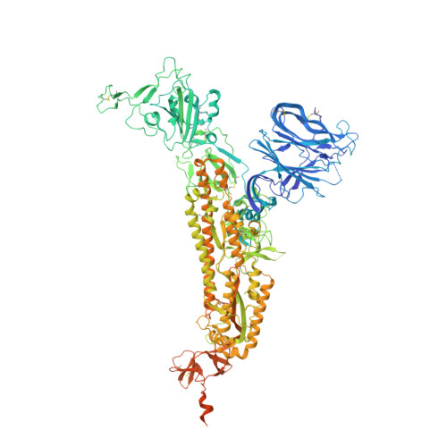
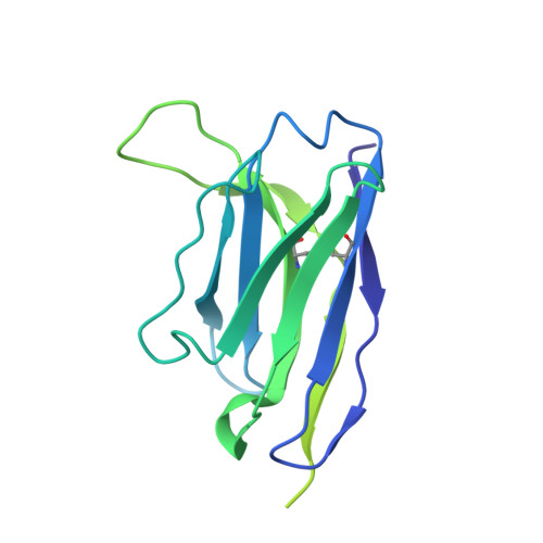
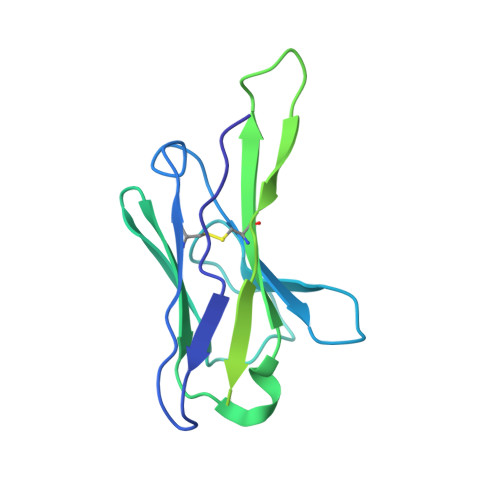
Potent SARS-CoV-2 neutralizing antibodies directed against spike N-terminal domain target a single supersite.

Cerutti, G.Guo, Y.Zhou, T.Gorman, J.Lee, M.Rapp, M.Reddem, E.R.Yu, J.Bahna, F.Bimela, J.Huang, Y.Katsamba, P.S.Liu, L.Nair, M.S.Rawi, R.Olia, A.S.Wang, P.Zhang, B.Chuang, G.Y.Ho, D.D.Sheng, Z.Kwong, P.D.Shapiro, L.

(2021) Cell Host Microbe 29: 819

  • DOI: https://doi.org/10.1016/j.chom.2021.03.005
  • Primary Citation Related Structures: 
    7L2C, 7L2D, 7L2E, 7L2F, 7LQW

  • PubMed Abstract: 

    Numerous antibodies that neutralize SARS-CoV-2 have been identified, and these generally target either the receptor-binding domain (RBD) or the N-terminal domain (NTD) of the viral spike. While RBD-directed antibodies have been extensively studied, far less is known about NTD-directed antibodies. Here, we report cryo-EM and crystal structures for seven potent NTD-directed neutralizing antibodies in complex with spike or isolated NTD. These structures defined several antibody classes, with at least one observed in multiple convalescent donors. The structures revealed that all seven antibodies target a common surface, bordered by glycans N17, N74, N122, and N149. This site-formed primarily by a mobile β-hairpin and several flexible loops-was highly electropositive, located at the periphery of the spike, and the largest glycan-free surface of NTD facing away from the viral membrane. Thus, in contrast to neutralizing RBD-directed antibodies that recognize multiple non-overlapping epitopes, potent NTD-directed neutralizing antibodies appear to target a single supersite.


  • Organizational Affiliation
    • Department of Biochemistry and Molecular Biophysics, Columbia University, New York, NY 10032, USA; Zuckerman Mind Brain Behavior Institute, Columbia University, New York, NY 10027, USA.

Macromolecule Content 

  • Total Structure Weight: 586.62 kDa 
  • Atom Count: 32,641 
  • Modeled Residue Count: 4,050 
  • Deposited Residue Count: 5,196 
  • Unique protein chains: 3

Macromolecules

Find similar proteins by:|  3D Structure
Entity ID: 1
MoleculeChains  Sequence LengthOrganismDetailsImage
Spike glycoprotein
A, B, C
1,288Severe acute respiratory syndrome coronavirus 2Mutation(s): 5 
Gene Names: S2
UniProt
Find proteins for P0DTC2 (Severe acute respiratory syndrome coronavirus 2)
Explore P0DTC2 
Go to UniProtKB:  P0DTC2
Entity Groups
Sequence Clusters30% Identity50% Identity70% Identity90% Identity95% Identity100% Identity
UniProt GroupP0DTC2
Glycosylation
Glycosylation Sites: 18Go to GlyGen: P0DTC2-1
Sequence Annotations
Expand
Reference Sequence
Find similar proteins by:|  3D Structure
Entity ID: 2
MoleculeChains  Sequence LengthOrganismDetailsImage
4-18 heavy chainD [auth H],
F [auth G],
H [auth I]
229Homo sapiensMutation(s): 0 
Find similar proteins by:|  3D Structure
Entity ID: 3
MoleculeChains  Sequence LengthOrganismDetailsImage
4-18 light chainE [auth L],
G [auth J],
I [auth K]
215Homo sapiensMutation(s): 0 

Small Molecules

Ligands 1 Unique
IDChains Name / Formula / InChI Key2D Diagram3D Interactions
NAG
(Subject of Investigation/LOI)

Query on NAG


Download:Ideal Coordinates CCD File
AB [auth B]
BB [auth C]
CB [auth C]
DB [auth C]
EB [auth C]
AB [auth B],
BB [auth C],
CB [auth C],
DB [auth C],
EB [auth C],
FB [auth C],
GB [auth C],
HA [auth A],
HB [auth C],
IA [auth A],
IB [auth C],
JA [auth A],
JB [auth C],
KA [auth A],
KB [auth C],
LA [auth A],
MA [auth A],
NA [auth A],
OA [auth A],
PA [auth A],
QA [auth A],
RA [auth B],
SA [auth B],
TA [auth B],
UA [auth B],
VA [auth B],
WA [auth B],
XA [auth B],
YA [auth B],
ZA [auth B]
2-acetamido-2-deoxy-beta-D-glucopyranose
C8 H15 N O6
OVRNDRQMDRJTHS-FMDGEEDCSA-N

Experimental Data & Validation

Experimental Data

  • Method: ELECTRON MICROSCOPY
  • Resolution: 2.97 Å
  • Aggregation State: PARTICLE 
  • Reconstruction Method: SINGLE PARTICLE 
EM Software:
TaskSoftware PackageVersion
RECONSTRUCTIONcryoSPARC

Structure Validation

View Full Validation Report


Entry History 

& Funding Information

Deposition Data


Funding OrganizationLocationGrant Number
Other privateChinaJack Ma Foundation

Revision History  (Full details and data files)

  • Version 1.0: 2021-03-24
    Type: Initial release
  • Version 1.1: 2021-04-14
    Changes: Database references
  • Version 1.2: 2021-05-26
    Changes: Database references
  • Version 1.3: 2024-11-06
    Changes: Data collection, Database references, Structure summary