7L1R | pdb_00007l1r

PS3 F1-ATPase Hydrolysis Dwell


Experimental Data Snapshot

  • Method: ELECTRON MICROSCOPY
  • Resolution: 3.10 Å
  • Aggregation State: PARTICLE 
  • Reconstruction Method: SINGLE PARTICLE 

wwPDB Validation 3D Report Full Report

Validation slider image for 7L1R

This is version 1.4 of the entry. See complete history

Literature

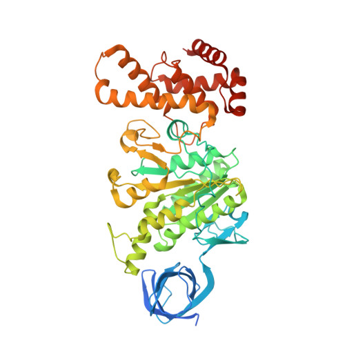
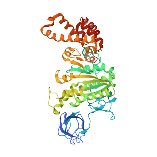
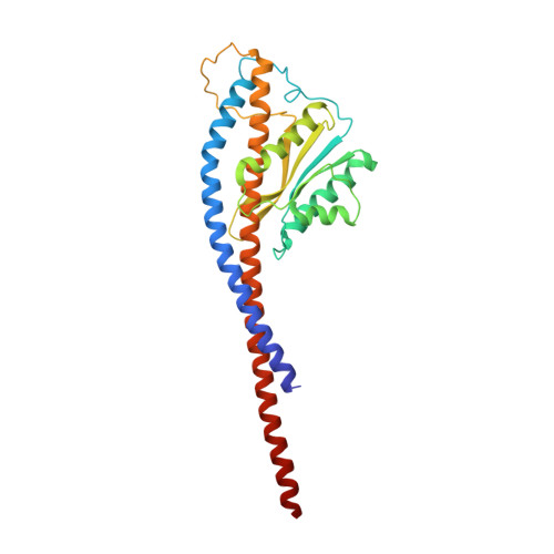
The six steps of the complete F 1 -ATPase rotary catalytic cycle.

Sobti, M.Ueno, H.Noji, H.Stewart, A.G.

(2021) Nat Commun 12: 4690-4690

  • DOI: https://doi.org/10.1038/s41467-021-25029-0
  • Primary Citation Related Structures: 
    7L1Q, 7L1R, 7L1S

  • PubMed Abstract: 

    F 1 F o ATP synthase interchanges phosphate transfer energy and proton motive force via a rotary catalysis mechanism. Isolated F 1 -ATPase catalytic cores can hydrolyze ATP, passing through six intermediate conformational states to generate rotation of their central γ-subunit. Although previous structural studies have contributed greatly to understanding rotary catalysis in the F 1 -ATPase, the structure of an important conformational state (the binding-dwell) has remained elusive. Here, we exploit temperature and time-resolved cryo-electron microscopy to determine the structure of the binding- and catalytic-dwell states of Bacillus PS3 F 1 -ATPase. Each state shows three catalytic β-subunits in different conformations, establishing the complete set of six states taken up during the catalytic cycle and providing molecular details for both the ATP binding and hydrolysis strokes. We also identify a potential phosphate-release tunnel that indicates how ADP and phosphate binding are coordinated during synthesis. Overall these findings provide a structural basis for the entire F 1 -ATPase catalytic cycle.


  • Organizational Affiliation
    • Molecular, Structural and Computational Biology Division, The Victor Chang Cardiac Research Institute, Darlinghurst, NSW, Australia.

Macromolecule Content 

  • Total Structure Weight: 362.85 kDa 
  • Atom Count: 24,199 
  • Modeled Residue Count: 3,118 
  • Deposited Residue Count: 3,267 
  • Unique protein chains: 3

Macromolecules

Find similar proteins by:|  3D Structure
Entity ID: 1
MoleculeChains  Sequence LengthOrganismDetailsImage
ATP synthase subunit alpha
A, B, C
510Bacillus sp. PS3Mutation(s): 0 
Gene Names: uncAatpA
EC: 7.1.2.2
Membrane Entity: Yes 
UniProt
Find proteins for A0A0M3VGF9 (Bacillus sp. (strain PS3))
Explore A0A0M3VGF9 
Go to UniProtKB:  A0A0M3VGF9
Entity Groups
Sequence Clusters30% Identity50% Identity70% Identity90% Identity95% Identity100% Identity
UniProt GroupA0A0M3VGF9
Sequence Annotations
Expand
Reference Sequence
Find similar proteins by:|  3D Structure
Entity ID: 2
MoleculeChains  Sequence LengthOrganismDetailsImage
ATP synthase subunit beta
D, E, F
484Bacillus sp. PS3Mutation(s): 0 
Gene Names: uncDatpD
EC: 7.1.2.2
Membrane Entity: Yes 
UniProt
Find proteins for P07677 (Bacillus sp. (strain PS3))
Explore P07677 
Go to UniProtKB:  P07677
Entity Groups
Sequence Clusters30% Identity50% Identity70% Identity90% Identity95% Identity100% Identity
UniProt GroupP07677
Sequence Annotations
Expand
Reference Sequence
Find similar proteins by:|  3D Structure
Entity ID: 3
MoleculeChains  Sequence LengthOrganismDetailsImage
ATP synthase gamma chain285Bacillus sp. PS3Mutation(s): 0 
Gene Names: uncGatpG
Membrane Entity: Yes 
UniProt
Find proteins for A0A0M4TPJ7 (Bacillus sp. (strain PS3))
Explore A0A0M4TPJ7 
Go to UniProtKB:  A0A0M4TPJ7
Entity Groups
Sequence Clusters30% Identity50% Identity70% Identity90% Identity95% Identity100% Identity
UniProt GroupA0A0M4TPJ7
Sequence Annotations
Expand
Reference Sequence

Small Molecules

Ligands 3 Unique
IDChains Name / Formula / InChI Key2D Diagram3D Interactions
ATP

Query on ATP


Download:Ideal Coordinates CCD File
H [auth A],
J [auth B],
L [auth C],
N [auth D],
R [auth F]
ADENOSINE-5'-TRIPHOSPHATE
C10 H16 N5 O13 P3
ZKHQWZAMYRWXGA-KQYNXXCUSA-N
ADP

Query on ADP


Download:Ideal Coordinates CCD File
P [auth E]ADENOSINE-5'-DIPHOSPHATE
C10 H15 N5 O10 P2
XTWYTFMLZFPYCI-KQYNXXCUSA-N
MG

Query on MG


Download:Ideal Coordinates CCD File
I [auth A]
K [auth B]
M [auth C]
O [auth D]
Q [auth E]
I [auth A],
K [auth B],
M [auth C],
O [auth D],
Q [auth E],
S [auth F]
MAGNESIUM ION
Mg
JLVVSXFLKOJNIY-UHFFFAOYSA-N

Experimental Data & Validation

Experimental Data

  • Method: ELECTRON MICROSCOPY
  • Resolution: 3.10 Å
  • Aggregation State: PARTICLE 
  • Reconstruction Method: SINGLE PARTICLE 
EM Software:
TaskSoftware PackageVersion
MODEL REFINEMENTPHENIX

Structure Validation

View Full Validation Report


Entry History 

Deposition Data

Revision History  (Full details and data files)

  • Version 1.0: 2021-07-21
    Type: Initial release
  • Version 1.1: 2021-08-25
    Changes: Database references
  • Version 1.2: 2021-09-01
    Changes: Database references
  • Version 1.3: 2024-05-29
    Changes: Data collection
  • Version 1.4: 2025-05-14
    Changes: Data collection, Structure summary