7KUH

MicroED structure of mVDAC


Experimental Data Snapshot

  • Method: ELECTRON CRYSTALLOGRAPHY
  • Resolution: 3.12 Å
  • R-Value Free: 0.287 
  • R-Value Work: 0.257 
  • R-Value Observed: 0.258 

wwPDB Validation   3D Report Full Report


This is version 1.2 of the entry. See complete history


Literature

MicroED structure of lipid-embedded mammalian mitochondrial voltage-dependent anion channel.

Martynowycz, M.W.Khan, F.Hattne, J.Abramson, J.Gonen, T.

(2020) Proc Natl Acad Sci U S A 117: 32380-32385

  • DOI: https://doi.org/10.1073/pnas.2020010117
  • Primary Citation of Related Structures:  
    7KUH

  • PubMed Abstract: 

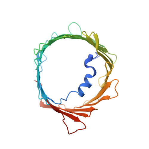
    A structure of the murine voltage-dependent anion channel (VDAC) was determined by microcrystal electron diffraction (MicroED). Microcrystals of an essential mutant of VDAC grew in a viscous bicelle suspension, making it unsuitable for conventional X-ray crystallography. Thin, plate-like crystals were identified using scanning-electron microscopy (SEM). Crystals were milled into thin lamellae using a focused-ion beam (FIB). MicroED data were collected from three crystal lamellae and merged for completeness. The refined structure revealed unmodeled densities between protein monomers, indicative of lipids that likely mediate contacts between the proteins in the crystal. This body of work demonstrates the effectiveness of milling membrane protein microcrystals grown in viscous media using a focused ion beam for subsequent structure determination by MicroED. This approach is well suited for samples that are intractable by X-ray crystallography. To our knowledge, the presented structure is a previously undescribed mutant of the membrane protein VDAC, crystallized in a lipid bicelle matrix and solved by MicroED.


  • Organizational Affiliation

    Howard Hughes Medical Institute, University of California, Los Angeles, CA 90095.


Macromolecules
Find similar proteins by:  (by identity cutoff)  |  3D Structure
Entity ID: 1
MoleculeChains Sequence LengthOrganismDetailsImage
Voltage-dependent anion-selective channel protein 1A [auth X]295Mus musculusMutation(s): 1 
Gene Names: Vdac1Vdac5
Membrane Entity: Yes 
UniProt & NIH Common Fund Data Resources
Find proteins for Q60932 (Mus musculus)
Explore Q60932 
Go to UniProtKB:  Q60932
IMPC:  MGI:106919
Entity Groups  
Sequence Clusters30% Identity50% Identity70% Identity90% Identity95% Identity100% Identity
UniProt GroupQ60932
Sequence Annotations
Expand
  • Reference Sequence
Experimental Data & Validation

Experimental Data

  • Method: ELECTRON CRYSTALLOGRAPHY
  • Resolution: 3.12 Å
  • R-Value Free: 0.287 
  • R-Value Work: 0.257 
  • R-Value Observed: 0.258 
  • Space Group: C 1 2 1
Unit Cell:
Length ( Å )Angle ( ˚ )
a = 98.9α = 90
b = 58.21β = 99.444
c = 69.54γ = 90
Software Package:
Software NamePurpose
phenix.refinerefinement
PHENIXrefinement
EM Software:
TaskSoftware PackageVersion
MODEL REFINEMENTPHENIX1.18.2
RECONSTRUCTIONPHENIX

Structure Validation

View Full Validation Report


Entry History & Funding Information

Deposition Data


Funding OrganizationLocationGrant Number
Howard Hughes Medical Institute (HHMI)United States--
National Institutes of Health/National Institute of General Medical Sciences (NIH/NIGMS)United StatesP41GM136508
National Institutes of Health/National Institute of General Medical Sciences (NIH/NIGMS)United StatesR35GM135175

Revision History  (Full details and data files)

  • Version 1.0: 2020-12-23
    Type: Initial release
  • Version 1.1: 2021-01-27
    Changes: Database references
  • Version 1.2: 2024-03-06
    Changes: Data collection, Database references, Refinement description