7KP0

CD1a-42:1 SM binary complex


Experimental Data Snapshot

  • Method: X-RAY DIFFRACTION
  • Resolution: 2.40 Å
  • R-Value Free: 0.264 
  • R-Value Work: 0.190 
  • R-Value Observed: 0.194 

wwPDB Validation   3D Report Full Report


Ligand Structure Quality Assessment 


This is version 1.2 of the entry. See complete history


Literature

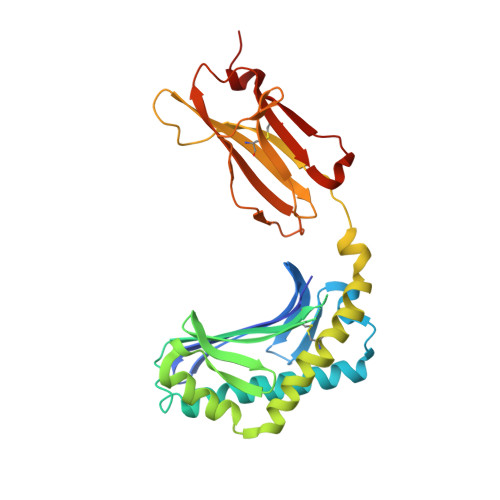
CD1a selectively captures endogenous cellular lipids that broadly block T cell response.

Cotton, R.N.Wegrecki, M.Cheng, T.Y.Chen, Y.L.Veerapen, N.Le Nours, J.Orgill, D.P.Pomahac, B.Talbot, S.G.Willis, R.Altman, J.D.de Jong, A.Van Rhijn, I.Clark, R.A.Besra, G.S.Ogg, G.Rossjohn, J.Moody, D.B.

(2021) J Exp Med 218

  • DOI: https://doi.org/10.1084/jem.20202699
  • Primary Citation of Related Structures:  
    7KOZ, 7KP0, 7KP1

  • PubMed Abstract: 

    We optimized lipidomics methods to broadly detect endogenous lipids bound to cellular CD1a proteins. Whereas membrane phospholipids dominate in cells, CD1a preferentially captured sphingolipids, especially a C42, doubly unsaturated sphingomyelin (42:2 SM). The natural 42:2 SM but not the more common 34:1 SM blocked CD1a tetramer binding to T cells in all human subjects tested. Thus, cellular CD1a selectively captures a particular endogenous lipid that broadly blocks its binding to TCRs. Crystal structures show that the short cellular SMs stabilized a triad of surface residues to remain flush with CD1a, but the longer lipids forced the phosphocholine group to ride above the display platform to hinder TCR approach. Whereas nearly all models emphasize antigen-mediated T cell activation, we propose that the CD1a system has intrinsic autoreactivity and is negatively regulated by natural endogenous inhibitors selectively bound in its cleft. Further, the detailed chemical structures of natural blockers could guide future design of therapeutic blockers of CD1a response.


  • Organizational Affiliation

    Graduate Program in Immunology, Harvard Medical School, Boston, MA.


Macromolecules
Find similar proteins by:  (by identity cutoff)  |  3D Structure
Entity ID: 1
MoleculeChains Sequence LengthOrganismDetailsImage
T-cell surface glycoprotein CD1a285Homo sapiensMutation(s): 0 
Gene Names: CD1A
UniProt & NIH Common Fund Data Resources
Find proteins for P06126 (Homo sapiens)
Explore P06126 
Go to UniProtKB:  P06126
PHAROS:  P06126
GTEx:  ENSG00000158477 
Entity Groups  
Sequence Clusters30% Identity50% Identity70% Identity90% Identity95% Identity100% Identity
UniProt GroupP06126
Sequence Annotations
Expand
  • Reference Sequence
Find similar proteins by:  (by identity cutoff)  |  3D Structure
Entity ID: 2
MoleculeChains Sequence LengthOrganismDetailsImage
Beta-2-microglobulin106Homo sapiensMutation(s): 0 
Gene Names: B2MCDABP0092HDCMA22P
UniProt & NIH Common Fund Data Resources
Find proteins for P61769 (Homo sapiens)
Explore P61769 
Go to UniProtKB:  P61769
PHAROS:  P61769
GTEx:  ENSG00000166710 
Entity Groups  
Sequence Clusters30% Identity50% Identity70% Identity90% Identity95% Identity100% Identity
UniProt GroupP61769
Sequence Annotations
Expand
  • Reference Sequence
Small Molecules
Ligands 2 Unique
IDChains Name / Formula / InChI Key2D Diagram3D Interactions
WUA (Subject of Investigation/LOI)
Query on WUA

Download Ideal Coordinates CCD File 
C [auth A](4R,7S)-4-hydroxy-7-[(1S,2E)-1-hydroxyhexadec-2-en-1-yl]-N,N,N-trimethyl-4,9-dioxo-3,5-dioxa-8-aza-4lambda~5~-phosphadotriacontan-1-aminium
C47 H96 N2 O6 P
QEDPUVGSSDPBMD-ZVBYXNGSSA-O
EDO
Query on EDO

Download Ideal Coordinates CCD File 
D [auth A],
E [auth A]
1,2-ETHANEDIOL
C2 H6 O2
LYCAIKOWRPUZTN-UHFFFAOYSA-N
Experimental Data & Validation

Experimental Data

  • Method: X-RAY DIFFRACTION
  • Resolution: 2.40 Å
  • R-Value Free: 0.264 
  • R-Value Work: 0.190 
  • R-Value Observed: 0.194 
  • Space Group: P 21 21 21
Unit Cell:
Length ( Å )Angle ( ˚ )
a = 42.469α = 90
b = 90.918β = 90
c = 107.351γ = 90
Software Package:
Software NamePurpose
PHENIXrefinement
XDSdata reduction
Aimlessdata scaling
PHASERphasing

Structure Validation

View Full Validation Report


Ligand Structure Quality Assessment 


Entry History 

Deposition Data

Revision History  (Full details and data files)

  • Version 1.0: 2021-05-05
    Type: Initial release
  • Version 1.1: 2021-06-16
    Changes: Database references
  • Version 1.2: 2023-10-18
    Changes: Data collection, Database references, Refinement description