7KNG

2.10A resolution structure of independent Phosphoglycerate mutase from C. elegans in complex with a macrocyclic peptide inhibitor (Ce-2 Y7F)


Experimental Data Snapshot

  • Method: X-RAY DIFFRACTION
  • Resolution: 2.10 Å
  • R-Value Free: 0.220 
  • R-Value Work: 0.167 
  • R-Value Observed: 0.169 

wwPDB Validation   3D Report Full Report


This is version 1.3 of the entry. See complete history


Literature

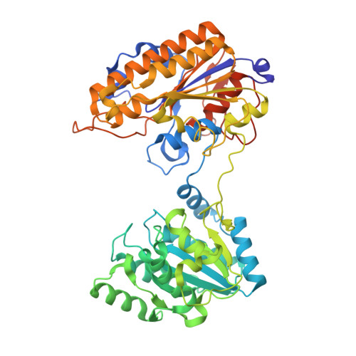
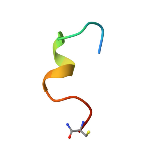
Structure-activity relationship of ipglycermide binding to phosphoglycerate mutases.

Wiedmann, M.Dranchak, P.K.Aitha, M.Queme, B.Collmus, C.D.Kashipathy, M.M.Kanter, L.Lamy, L.Rogers, J.M.Tao, D.Battaile, K.P.Rai, G.Lovell, S.Suga, H.Inglese, J.

(2021) J Biol Chem 296: 100628-100628

  • DOI: https://doi.org/10.1016/j.jbc.2021.100628
  • Primary Citation of Related Structures:  
    7KNF, 7KNG

  • PubMed Abstract: 

    Catalysis of human phosphoglycerate mutase is dependent on a 2,3-bisphosphoglycerate cofactor (dPGM), whereas the nonhomologous isozyme in many parasitic species is cofactor independent (iPGM). This mechanistic and phylogenetic diversity offers an opportunity for selective pharmacologic targeting of glycolysis in disease-causing organisms. We previously discovered ipglycermide, a potent inhibitor of iPGM, from a large combinatorial cyclic peptide library. To fully delineate the ipglycermide pharmacophore, herein we construct a detailed structure-activity relationship using 280 substituted ipglycermide analogs. Binding affinities of these analogs to immobilized Caenorhabditis elegans iPGM, measured as fold enrichment relative to the index residue by deep sequencing of an mRNA display library, illuminated the significance of each amino acid to the pharmacophore. Using cocrystal structures and binding kinetics, we show that the high affinity of ipglycermide for iPGM orthologs, from Brugia malayi, Onchocerca volvulus, Dirofilaria immitis, and Escherichia coli, is achieved by a codependence between (1) the off-rate mediated by the macrocycle Cys14 thiolate coordination to an active-site Zn 2+ in the iPGM phosphatase domain and (2) shape complementarity surrounding the macrocyclic core at the phosphotransferase-phosphatase domain interface. Our results show that the high-affinity binding of ipglycermide to iPGMs freezes these structurally dynamic enzymes into an inactive, stable complex.


  • Organizational Affiliation

    Department of Chemistry, Graduate School of Sciences, The University of Tokyo, Tokyo, Japan.


Macromolecules
Find similar proteins by:  (by identity cutoff)  |  3D Structure
Entity ID: 1
MoleculeChains Sequence LengthOrganismDetailsImage
2,3-bisphosphoglycerate-independent phosphoglycerate mutase
A, B
538Caenorhabditis elegansMutation(s): 0 
Gene Names: ipgm-1F57B10.3
EC: 5.4.2.12
UniProt
Find proteins for G5EFZ1 (Caenorhabditis elegans)
Explore G5EFZ1 
Go to UniProtKB:  G5EFZ1
Entity Groups  
Sequence Clusters30% Identity50% Identity70% Identity90% Identity95% Identity100% Identity
UniProt GroupG5EFZ1
Sequence Annotations
Expand
  • Reference Sequence

Find similar proteins by:  Sequence   |   3D Structure  

Entity ID: 2
MoleculeChains Sequence LengthOrganismDetailsImage
DTY-ASP-TYR-PRO-GLY-ASP-PHE-CYS-TYR-LEU-TYR-GLY-THR-CYS
C, D
16Caenorhabditis elegansMutation(s): 0 
Entity Groups  
Sequence Clusters30% Identity50% Identity70% Identity90% Identity95% Identity100% Identity
Sequence Annotations
Expand
  • Reference Sequence
Small Molecules
Ligands 3 Unique
IDChains Name / Formula / InChI Key2D Diagram3D Interactions
ZN
Query on ZN

Download Ideal Coordinates CCD File 
I [auth A],
J [auth A],
O [auth B],
P [auth B]
ZINC ION
Zn
PTFCDOFLOPIGGS-UHFFFAOYSA-N
CL
Query on CL

Download Ideal Coordinates CCD File 
E [auth A],
F [auth A],
K [auth B],
L [auth B]
CHLORIDE ION
Cl
VEXZGXHMUGYJMC-UHFFFAOYSA-M
NA
Query on NA

Download Ideal Coordinates CCD File 
G [auth A],
H [auth A],
M [auth B],
N [auth B]
SODIUM ION
Na
FKNQFGJONOIPTF-UHFFFAOYSA-N
Experimental Data & Validation

Experimental Data

  • Method: X-RAY DIFFRACTION
  • Resolution: 2.10 Å
  • R-Value Free: 0.220 
  • R-Value Work: 0.167 
  • R-Value Observed: 0.169 
  • Space Group: P 1 2 1
Unit Cell:
Length ( Å )Angle ( ˚ )
a = 73.862α = 90
b = 75.471β = 99.11
c = 101.36γ = 90
Software Package:
Software NamePurpose
XDSdata reduction
Aimlessdata scaling
PHASERphasing
PHENIXrefinement
PDB_EXTRACTdata extraction

Structure Validation

View Full Validation Report


Entry History & Funding Information

Deposition Data


Funding OrganizationLocationGrant Number
National Institutes of Health/National Center for Advancing Translational Sciences (NIH/NCATS)United States1ZIATR000247
National Institutes of Health/National Institute of General Medical Sciences (NIH/NIGMS)United StatesP30GM110761

Revision History  (Full details and data files)

  • Version 1.0: 2021-04-07
    Type: Initial release
  • Version 1.1: 2021-04-21
    Changes: Database references
  • Version 1.2: 2021-07-14
    Changes: Database references
  • Version 1.3: 2023-10-18
    Changes: Data collection, Database references, Refinement description