Structural and functional analyses explain Pea KAI2 receptor diversity and reveal stereoselective catalysis during signal perception.
Guercio, A.M., Torabi, S., Cornu, D., Dalmais, M., Bendahmane, A., Le Signor, C., Pillot, J.P., Le Bris, P., Boyer, F.D., Rameau, C., Gutjahr, C., de Saint Germain, A., Shabek, N.(2022) Commun Biol 5: 126-126
- PubMed: 35149763 Search on PubMedSearch on PubMed Central
- DOI: https://doi.org/10.1038/s42003-022-03085-6
- Primary Citation Related Structures:
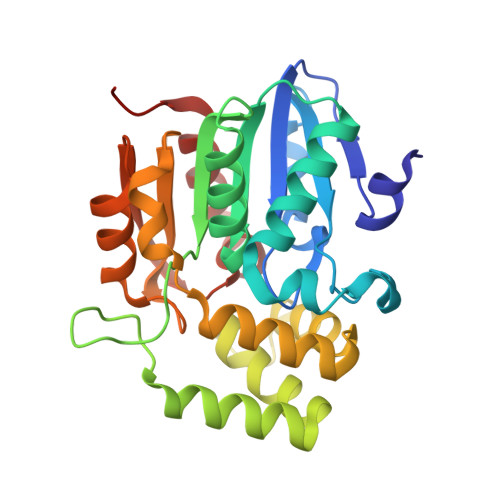
7K2Z, 7K38 - PubMed Abstract:
KAI2 proteins are plant α/β hydrolase receptors which perceive smoke-derived butenolide signals and endogenous, yet unidentified KAI2-ligands (KLs). The number of functional KAI2 receptors varies among species and KAI2 gene duplication and sub-functionalization likely plays an adaptative role by altering specificity towards different KLs. Legumes represent one of the largest families of flowering plants and contain many agronomic crops. Prior to their diversification, KAI2 underwent duplication resulting in KAI2A and KAI2B. Here we demonstrate that Pisum sativum KAI2A and KAI2B are active receptors and enzymes with divergent ligand stereoselectivity. KAI2B has a higher affinity for and hydrolyses a broader range of substrates including strigolactone-like stereoisomers. We determine the crystal structures of PsKAI2B in apo and butenolide-bound states. The biochemical, structural, and mass spectra analyses of KAI2s reveal a transient intermediate on the catalytic serine and a stable adduct on the catalytic histidine, confirming its role as a bona fide enzyme. Our work uncovers the stereoselectivity of ligand perception and catalysis by diverged KAI2 receptors and proposes adaptive sensitivity to KAR/KL and strigolactones by KAI2B.
- Department of Plant Biology, College of Biological Sciences, University of California, Davis, CA, 95616, USA.
Organizational Affiliation: