7JZL | pdb_00007jzl

SARS-CoV-2 spike in complex with LCB1 (2RBDs open)


Experimental Data Snapshot

  • Method: ELECTRON MICROSCOPY
  • Resolution: 2.70 Å
  • Aggregation State: PARTICLE 
  • Reconstruction Method: SINGLE PARTICLE 

wwPDB Validation 3D Report Full Report

Validation slider image for 7JZL

This is version 1.2 of the entry. See complete history

Literature

De novo design of picomolar SARS-CoV-2 miniprotein inhibitors.

Cao, L.Goreshnik, I.Coventry, B.Case, J.B.Miller, L.Kozodoy, L.Chen, R.E.Carter, L.Walls, A.C.Park, Y.J.Strauch, E.M.Stewart, L.Diamond, M.S.Veesler, D.Baker, D.

(2020) Science 370: 426-431

  • DOI: https://doi.org/10.1126/science.abd9909
  • Primary Citation Related Structures: 
    7JZL, 7JZM, 7JZN, 7JZU

  • PubMed Abstract: 

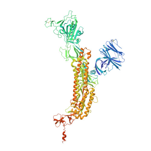
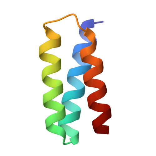
    Targeting the interaction between the severe acute respiratory syndrome coronavirus 2 (SARS-CoV-2) spike protein and the human angiotensin-converting enzyme 2 (ACE2) receptor is a promising therapeutic strategy. We designed inhibitors using two de novo design approaches. Computer-generated scaffolds were either built around an ACE2 helix that interacts with the spike receptor binding domain (RBD) or docked against the RBD to identify new binding modes, and their amino acid sequences were designed to optimize target binding, folding, and stability. Ten designs bound the RBD, with affinities ranging from 100 picomolar to 10 nanomolar, and blocked SARS-CoV-2 infection of Vero E6 cells with median inhibitory concentration (IC 50 ) values between 24 picomolar and 35 nanomolar. The most potent, with new binding modes, are 56- and 64-residue proteins (IC 50 ~ 0.16 nanograms per milliliter). Cryo-electron microscopy structures of these minibinders in complex with the SARS-CoV-2 spike ectodomain trimer with all three RBDs bound are nearly identical to the computational models. These hyperstable minibinders provide starting points for SARS-CoV-2 therapeutics.


  • Organizational Affiliation
    • Department of Biochemistry, University of Washington, Seattle, WA 98195, USA.

Macromolecule Content 

  • Total Structure Weight: 460.09 kDa 
  • Atom Count: 23,123 
  • Modeled Residue Count: 3,105 
  • Deposited Residue Count: 4,029 
  • Unique protein chains: 2

Macromolecules

Find similar proteins by:|  3D Structure
Entity ID: 1
MoleculeChains  Sequence LengthOrganismDetailsImage
Spike glycoproteinA,
C,
E [auth B]
1,288Severe acute respiratory syndrome coronavirus 2Mutation(s): 0 
Gene Names: S2
UniProt
Find proteins for P0DTC2 (Severe acute respiratory syndrome coronavirus 2)
Explore P0DTC2 
Go to UniProtKB:  P0DTC2
Entity Groups
Sequence Clusters30% Identity50% Identity70% Identity90% Identity95% Identity100% Identity
UniProt GroupP0DTC2
Glycosylation
Glycosylation Sites: 16Go to GlyGen: P0DTC2-1
Sequence Annotations
Expand
Reference Sequence
Find similar proteins by:|  3D Structure
Entity ID: 2
MoleculeChains  Sequence LengthOrganismDetailsImage
LCB1B [auth E],
D [auth G],
F
55synthetic constructMutation(s): 0 

Oligosaccharides

Help  
Entity ID: 3
MoleculeChains Length2D Diagram GlycosylationD Interactions
2-acetamido-2-deoxy-beta-D-glucopyranose-(1-4)-2-acetamido-2-deoxy-beta-D-glucopyranose
G [auth D],
H,
I,
J,
K,
G [auth D],
H,
I,
J,
K,
L,
M,
N,
O,
P,
Q,
R,
S
2N-Glycosylation
Glycosylation Resources
GlyTouCan: G42666HT
GlyCosmos: G42666HT
GlyGen: G42666HT

Small Molecules

Ligands 1 Unique
IDChains Name / Formula / InChI Key2D Diagram3D Interactions
NAG

Query on NAG


Download:Ideal Coordinates CCD File
AA [auth A]
BA [auth A]
CA [auth A]
DA [auth A]
EA [auth C]
AA [auth A],
BA [auth A],
CA [auth A],
DA [auth A],
EA [auth C],
FA [auth C],
GA [auth C],
HA [auth C],
IA [auth C],
JA [auth C],
KA [auth C],
LA [auth C],
MA [auth C],
NA [auth C],
OA [auth C],
PA [auth B],
QA [auth B],
RA [auth B],
SA [auth B],
T [auth A],
TA [auth B],
U [auth A],
UA [auth B],
V [auth A],
VA [auth B],
W [auth A],
WA [auth B],
X [auth A],
XA [auth B],
Y [auth A],
YA [auth B],
Z [auth A],
ZA [auth B]
2-acetamido-2-deoxy-beta-D-glucopyranose
C8 H15 N O6
OVRNDRQMDRJTHS-FMDGEEDCSA-N

Experimental Data & Validation

Experimental Data

  • Method: ELECTRON MICROSCOPY
  • Resolution: 2.70 Å
  • Aggregation State: PARTICLE 
  • Reconstruction Method: SINGLE PARTICLE 
EM Software:
TaskSoftware PackageVersion
RECONSTRUCTIONcryoSPARC

Structure Validation

View Full Validation Report


Entry History 

& Funding Information

Deposition Data


Funding OrganizationLocationGrant Number
National Institutes of Health/National Institute of General Medical Sciences (NIH/NIGMS)United StatesR01GM120553

Revision History  (Full details and data files)

  • Version 1.0: 2020-09-23
    Type: Initial release
  • Version 1.1: 2020-11-04
    Changes: Database references
  • Version 1.2: 2024-10-30
    Changes: Data collection, Database references, Structure summary