7JVB | pdb_00007jvb

Crystal structure of the SARS-CoV-2 spike receptor-binding domain (RBD) with nanobody Nb20


Experimental Data Snapshot

  • Method: X-RAY DIFFRACTION
  • Resolution: 3.29 Å
  • R-Value Free: 
    0.322 (Depositor), 0.311 (DCC) 
  • R-Value Work: 
    0.281 (Depositor), 0.284 (DCC) 
  • R-Value Observed: 
    0.283 (Depositor) 

Starting Model: experimental
View more details

wwPDB Validation 3D Report Full Report

Validation slider image for 7JVB

This is version 1.4 of the entry. See complete history

Literature

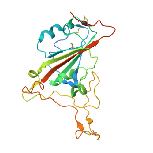
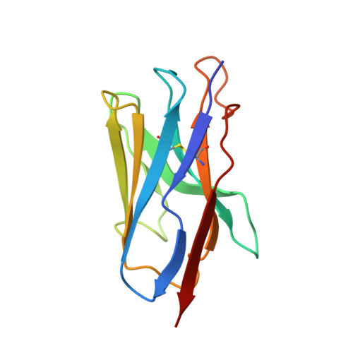
Versatile and multivalent nanobodies efficiently neutralize SARS-CoV-2.

Xiang, Y.Nambulli, S.Xiao, Z.Liu, H.Sang, Z.Duprex, W.P.Schneidman-Duhovny, D.Zhang, C.Shi, Y.

(2020) Science 370: 1479-1484

  • DOI: https://doi.org/10.1126/science.abe4747
  • Primary Citation Related Structures: 
    7JVB

  • PubMed Abstract: 

    Cost-effective, efficacious therapeutics are urgently needed to combat the COVID-19 pandemic. In this study, we used camelid immunization and proteomics to identify a large repertoire of highly potent neutralizing nanobodies (Nbs) to the severe acute respiratory syndrome coronavirus 2 (SARS-CoV-2) spike protein receptor binding domain (RBD). We discovered Nbs with picomolar to femtomolar affinities that inhibit viral infection at concentrations below the nanograms-per-milliliter level, and we determined a structure of one of the most potent Nbs in complex with the RBD. Structural proteomics and integrative modeling revealed multiple distinct and nonoverlapping epitopes and indicated an array of potential neutralization mechanisms. We bioengineered multivalent Nb constructs that achieved ultrahigh neutralization potency (half-maximal inhibitory concentration as low as 0.058 ng/ml) and may prevent mutational escape. These thermostable Nbs can be rapidly produced in bulk from microbes and resist lyophilization and aerosolization.


  • Organizational Affiliation
    • Department of Cell Biology, University of Pittsburgh, Pittsburgh, PA, USA.

Macromolecule Content 

  • Total Structure Weight: 76.01 kDa 
  • Atom Count: 4,748 
  • Modeled Residue Count: 621 
  • Deposited Residue Count: 682 
  • Unique protein chains: 2

Macromolecules

Find similar proteins by:|  3D Structure
Entity ID: 1
MoleculeChains  Sequence LengthOrganismDetailsImage
Spike protein S1A,
C [auth B]
224Severe acute respiratory syndrome coronavirus 2Mutation(s): 0 
Gene Names: S2
UniProt
Find proteins for P0DTC2 (Severe acute respiratory syndrome coronavirus 2)
Explore P0DTC2 
Go to UniProtKB:  P0DTC2
Entity Groups
Sequence Clusters30% Identity50% Identity70% Identity90% Identity95% Identity100% Identity
UniProt GroupP0DTC2
Sequence Annotations
Expand
Reference Sequence
Find similar proteins by:|  3D Structure
Entity ID: 2
MoleculeChains  Sequence LengthOrganismDetailsImage
Nanobody Nb20B [auth C],
D
117Lama glamaMutation(s): 0 

Experimental Data & Validation

Experimental Data

  • Method: X-RAY DIFFRACTION
  • Resolution: 3.29 Å
  • R-Value Free:  0.322 (Depositor), 0.311 (DCC) 
  • R-Value Work:  0.281 (Depositor), 0.284 (DCC) 
  • R-Value Observed: 0.283 (Depositor) 
Space Group: P 41 21 2
Unit Cell:
Length ( Å )Angle ( ˚ )
a = 70.716α = 90
b = 70.716β = 90
c = 435.037γ = 90
Software Package:
Software NamePurpose
PHENIXrefinement
JBluIce-EPICSdata collection
HKL-2000data reduction
HKL-2000data scaling
PHENIXphasing

Structure Validation

View Full Validation Report


Entry History 

& Funding Information

Deposition Data


Funding OrganizationLocationGrant Number
National Institutes of Health/National Institute of General Medical Sciences (NIH/NIGMS)United StatesR35GM128641

Revision History  (Full details and data files)

  • Version 1.0: 2020-12-02
    Type: Initial release
  • Version 1.1: 2020-12-30
    Changes: Database references
  • Version 1.2: 2021-01-27
    Changes: Structure summary
  • Version 1.3: 2023-10-18
    Changes: Data collection, Database references, Refinement description
  • Version 1.4: 2024-11-20
    Changes: Structure summary