Investigating the reaction and substrate preference of indole-3-acetaldehyde dehydrogenase from the plant pathogen Pseudomonas syringae PtoDC3000.
Zhang, K., Lee, J.S., Liu, R., Chan, Z.T., Dawson, T.J., De Togni, E.S., Edwards, C.T., Eng, I.K., Gao, A.R., Goicouria, L.A., Hall, E.M., Hu, K.A., Huang, K., Kizhner, A., Kodama, K.C., Lin, A.Z., Liu, J.Y., Lu, A.Y., Peng, O.W., Ryu, E.P., Shi, S., Sorkin, M.L., Walker, P.L., Wang, G.J., Xu, M.C., Yang, R.S., Cascella, B., Cruz, W., Holland, C.K., McClerkin, S.A., Kunkel, B.N., Lee, S.G., Jez, J.M.(2020) Biosci Rep 40
- PubMed: 33325526 Search on PubMedSearch on PubMed Central
- DOI: https://doi.org/10.1042/BSR20202959
- Primary Citation Related Structures:
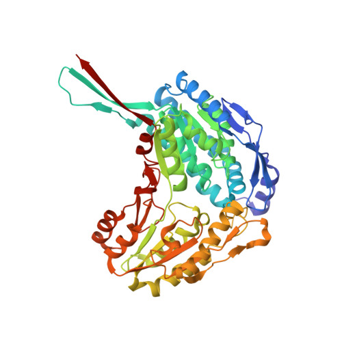
7JSO - PubMed Abstract:
Aldehyde dehydrogenases (ALDHs) catalyze the conversion of various aliphatic and aromatic aldehydes into corresponding carboxylic acids. Traditionally considered as housekeeping enzymes, new biochemical roles are being identified for members of ALDH family. Recent work showed that AldA from the plant pathogen Pseudomonas syringae strain PtoDC3000 (PtoDC3000) functions as an indole-3-acetaldehyde dehydrogenase for the synthesis of indole-3-acetic acid (IAA). IAA produced by AldA allows the pathogen to suppress salicylic acid-mediated defenses in the model plant Arabidopsis thaliana. Here we present a biochemical and structural analysis of the AldA indole-3-acetaldehyde dehydrogenase from PtoDC3000. Site-directed mutants targeting the catalytic residues Cys302 and Glu267 resulted in a loss of enzymatic activity. The X-ray crystal structure of the catalytically inactive AldA C302A mutant in complex with IAA and NAD+ showed the cofactor adopting a conformation that differs from the previously reported structure of AldA. These structures suggest that NAD+ undergoes a conformational change during the AldA reaction mechanism similar to that reported for human ALDH. Site-directed mutagenesis of the IAA binding site indicates that changes in the active site surface reduces AldA activity; however, substitution of Phe169 with a tryptophan altered the substrate selectivity of the mutant to prefer octanal. The present study highlights the inherent biochemical versatility of members of the ALDH enzyme superfamily in P. syringae.
- Department of Biology, Washington University in St. Louis, St. Louis, MO 63130, U.S.A.
Organizational Affiliation: