7JQ7

The Phi-28 gp11 DNA packaging Motor


Experimental Data Snapshot

  • Method: X-RAY DIFFRACTION
  • Resolution: 2.90 Å
  • R-Value Free: 0.284 
  • R-Value Work: 0.235 
  • R-Value Observed: 0.237 

wwPDB Validation   3D Report Full Report


This is version 1.3 of the entry. See complete history


Literature

Atomistic basis of force generation, translocation, and coordination in a viral genome packaging motor.

Pajak, J.Dill, E.Reyes-Aldrete, E.White, M.A.Kelch, B.A.Jardine, P.J.Arya, G.Morais, M.C.

(2021) Nucleic Acids Res 49: 6474-6488

  • DOI: https://doi.org/10.1093/nar/gkab372
  • Primary Citation of Related Structures:  
    7JQ6, 7JQ7, 7JQP

  • PubMed Abstract: 

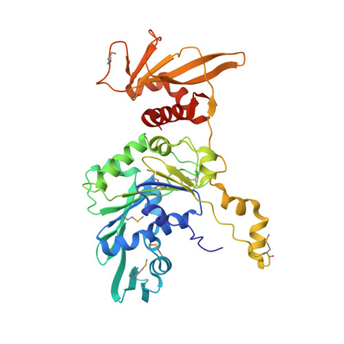
    Double-stranded DNA viruses package their genomes into pre-assembled capsids using virally-encoded ASCE ATPase ring motors. We present the first atomic-resolution crystal structure of a multimeric ring form of a viral dsDNA packaging motor, the ATPase of the asccφ28 phage, and characterize its atomic-level dynamics via long timescale molecular dynamics simulations. Based on these results, and previous single-molecule data and cryo-EM reconstruction of the homologous φ29 motor, we propose an overall packaging model that is driven by helical-to-planar transitions of the ring motor. These transitions are coordinated by inter-subunit interactions that regulate catalytic and force-generating events. Stepwise ATP binding to individual subunits increase their affinity for the helical DNA phosphate backbone, resulting in distortion away from the planar ring towards a helical configuration, inducing mechanical strain. Subsequent sequential hydrolysis events alleviate the accumulated mechanical strain, allowing a stepwise return of the motor to the planar conformation, translocating DNA in the process. This type of helical-to-planar mechanism could serve as a general framework for ring ATPases.


  • Organizational Affiliation

    Dept. of Mechanical Engineering and Materials Science, Duke University, Durham, NC 27708, USA.


Macromolecules
Find similar proteins by:  (by identity cutoff)  |  3D Structure
Entity ID: 1
MoleculeChains Sequence LengthOrganismDetailsImage
Encapsidation protein
A, B, C, D, E
374Lactococcus phage asccphi28Mutation(s): 0 
UniProt
Find proteins for B1ABI1 (Lactococcus phage asccphi28)
Explore B1ABI1 
Go to UniProtKB:  B1ABI1
Entity Groups  
Sequence Clusters30% Identity50% Identity70% Identity90% Identity95% Identity100% Identity
UniProt GroupB1ABI1
Sequence Annotations
Expand
  • Reference Sequence
Small Molecules
Ligands 2 Unique
IDChains Name / Formula / InChI Key2D Diagram3D Interactions
IOD
Query on IOD

Download Ideal Coordinates CCD File 
AA [auth A]
AB [auth B]
AC [auth C]
AD [auth D]
AE [auth E]
AA [auth A],
AB [auth B],
AC [auth C],
AD [auth D],
AE [auth E],
BA [auth A],
BB [auth B],
BC [auth C],
BD [auth D],
BE [auth E],
CA [auth A],
CB [auth B],
CC [auth C],
CD [auth D],
CE [auth E],
DA [auth A],
DB [auth B],
DC [auth C],
DD [auth D],
DE [auth E],
EA [auth A],
EB [auth B],
EC [auth C],
ED [auth D],
EE [auth E],
F [auth A],
FA [auth A],
FB [auth B],
FC [auth C],
FD [auth D],
FE [auth E],
G [auth A],
GA [auth A],
GB [auth B],
GC [auth C],
GD [auth D],
GE [auth E],
H [auth A],
HA [auth A],
HB [auth B],
HC [auth C],
HD [auth D],
I [auth A],
IA [auth B],
IB [auth B],
IC [auth C],
ID [auth D],
J [auth A],
JA [auth B],
JB [auth B],
JC [auth C],
JD [auth D],
K [auth A],
KA [auth B],
KB [auth C],
KC [auth C],
KD [auth D],
L [auth A],
LA [auth B],
LB [auth C],
LC [auth C],
LD [auth D],
M [auth A],
MA [auth B],
MB [auth C],
MC [auth C],
MD [auth E],
N [auth A],
NA [auth B],
NB [auth C],
NC [auth C],
ND [auth E],
O [auth A],
OA [auth B],
OB [auth C],
OC [auth C],
OD [auth E],
P [auth A],
PA [auth B],
PB [auth C],
PC [auth C],
PD [auth E],
Q [auth A],
QA [auth B],
QB [auth C],
QC [auth C],
QD [auth E],
R [auth A],
RA [auth B],
RB [auth C],
RC [auth C],
RD [auth E],
S [auth A],
SA [auth B],
SB [auth C],
SC [auth C],
SD [auth E],
T [auth A],
TA [auth B],
TB [auth C],
TD [auth E],
U [auth A],
UA [auth B],
UB [auth C],
UC [auth D],
UD [auth E],
V [auth A],
VA [auth B],
VB [auth C],
VC [auth D],
VD [auth E],
W [auth A],
WA [auth B],
WB [auth C],
WC [auth D],
WD [auth E],
X [auth A],
XA [auth B],
XB [auth C],
XC [auth D],
XD [auth E],
Y [auth A],
YA [auth B],
YB [auth C],
YC [auth D],
YD [auth E],
Z [auth A],
ZA [auth B],
ZB [auth C],
ZC [auth D],
ZD [auth E]
IODIDE ION
I
XMBWDFGMSWQBCA-UHFFFAOYSA-M
SO4
Query on SO4

Download Ideal Coordinates CCD File 
HE [auth E],
TC [auth C]
SULFATE ION
O4 S
QAOWNCQODCNURD-UHFFFAOYSA-L
Modified Residues  1 Unique
IDChains TypeFormula2D DiagramParent
MSE
Query on MSE
A, B, C, D, E
L-PEPTIDE LINKINGC5 H11 N O2 SeMET
Experimental Data & Validation

Experimental Data

  • Method: X-RAY DIFFRACTION
  • Resolution: 2.90 Å
  • R-Value Free: 0.284 
  • R-Value Work: 0.235 
  • R-Value Observed: 0.237 
  • Space Group: P 43 21 2
Unit Cell:
Length ( Å )Angle ( ˚ )
a = 112.2α = 90
b = 112.2β = 90
c = 354.2γ = 90
Software Package:
Software NamePurpose
HKL-2000data scaling
PHASERphasing
PHENIXrefinement
PDB_EXTRACTdata extraction
HKL-2000data reduction

Structure Validation

View Full Validation Report


Entry History & Funding Information

Deposition Data


Funding OrganizationLocationGrant Number
National Institutes of Health/National Institute of General Medical Sciences (NIH/NIGMS)United StatesGM095516
National Institutes of Health/National Institute of General Medical Sciences (NIH/NIGMS)United StatesGM122979

Revision History  (Full details and data files)

  • Version 1.0: 2021-06-16
    Type: Initial release
  • Version 1.1: 2021-07-14
    Changes: Database references
  • Version 1.2: 2023-10-18
    Changes: Data collection, Database references, Refinement description
  • Version 1.3: 2023-11-15
    Changes: Data collection