7JMU

Hen egg-white lysozyme with ionic liquid ethylammonium nitrate


Experimental Data Snapshot

  • Method: X-RAY DIFFRACTION
  • Resolution: 1.20 Å
  • R-Value Free: 0.183 
  • R-Value Work: 0.147 
  • R-Value Observed: 0.149 

wwPDB Validation   3D Report Full Report


This is version 1.2 of the entry. See complete history


Literature

Lysozyme conformational changes with ionic liquids: Spectroscopic, small angle x-ray scattering and crystallographic study.

Han, Q.Smith, K.M.Darmanin, C.Ryan, T.M.Drummond, C.J.Greaves, T.L.

(2021) J Colloid Interface Sci 585: 433-443

  • DOI: https://doi.org/10.1016/j.jcis.2020.10.024
  • Primary Citation of Related Structures:  
    7JMU

  • PubMed Abstract: 

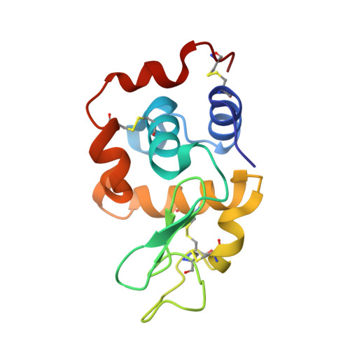
    Solvents that support protein functionality are important for biochemical applications, and new solvents are required. Here we employ FTIR and fluorescence spectroscopies, small angle X-ray scattering (SAXS) and X-ray crystallography to understand conformational changes of lysozyme with ionic liquids (ILs) added. Spectroscopic techniques identified that the secondary structure of lysozyme was maintained at the lower IL concentrations of 1 and 5 mol%, though the Tryptophan environment was significantly altered with nitrate-based ILs present. SAXS experiments indicated that the radius of gyration of lysozyme increased with 1 mol% IL present, and then decreased with increasing IL concentrations. The tertiary structure, particularly the loop regions, changed as a function of IL concentration, and this depended on the IL type. The crystallographic structure of lysozyme with the IL of ethylammonium nitrate present confirmed the loop region was extended, and identified three specific binding sites with nitrate ions, and that the positively charged areas were IL sensitive regions. This work provides a detailed understanding of lysozyme conformational changes in the presence of ILs. This approach can be extended to other functionally-important proteins.


  • Organizational Affiliation

    School of Science, College of Science, Engineering and Health, RMIT University, 124 La Trobe Street, Melbourne, VIC 3000, Australia.


Macromolecules
Find similar proteins by:  (by identity cutoff)  |  3D Structure
Entity ID: 1
MoleculeChains Sequence LengthOrganismDetailsImage
Lysozyme C
A, B
129Gallus gallusMutation(s): 0 
EC: 3.2.1.17
UniProt
Find proteins for P00698 (Gallus gallus)
Explore P00698 
Go to UniProtKB:  P00698
Entity Groups  
Sequence Clusters30% Identity50% Identity70% Identity90% Identity95% Identity100% Identity
UniProt GroupP00698
Sequence Annotations
Expand
  • Reference Sequence
Small Molecules
Ligands 1 Unique
IDChains Name / Formula / InChI Key2D Diagram3D Interactions
NO3 (Subject of Investigation/LOI)
Query on NO3

Download Ideal Coordinates CCD File 
C [auth A]
D [auth A]
E [auth A]
F [auth A]
G [auth A]
C [auth A],
D [auth A],
E [auth A],
F [auth A],
G [auth A],
H [auth A],
I [auth A],
J [auth B],
K [auth B],
L [auth B],
M [auth B],
N [auth B],
O [auth B],
P [auth B],
Q [auth B]
NITRATE ION
N O3
NHNBFGGVMKEFGY-UHFFFAOYSA-N
Experimental Data & Validation

Experimental Data

  • Method: X-RAY DIFFRACTION
  • Resolution: 1.20 Å
  • R-Value Free: 0.183 
  • R-Value Work: 0.147 
  • R-Value Observed: 0.149 
  • Space Group: P 1 21 1
Unit Cell:
Length ( Å )Angle ( ˚ )
a = 27.585α = 90
b = 62.637β = 90.54
c = 59.645γ = 90
Software Package:
Software NamePurpose
REFMACrefinement
Aimlessdata scaling
PDB_EXTRACTdata extraction
XDSdata reduction
PHENIXphasing

Structure Validation

View Full Validation Report


Entry History 

Deposition Data

Revision History  (Full details and data files)

  • Version 1.0: 2020-11-11
    Type: Initial release
  • Version 1.1: 2021-01-13
    Changes: Database references
  • Version 1.2: 2023-10-18
    Changes: Data collection, Database references, Refinement description