Structural basis of KdpD histidine kinase binding to the second messenger c-di-AMP.
Dutta, A., Batish, M., Parashar, V.(2021) J Biol Chem 296: 100771-100771
- PubMed: 33989637 Search on PubMedSearch on PubMed Central
- DOI: https://doi.org/10.1016/j.jbc.2021.100771
- Primary Citation Related Structures:
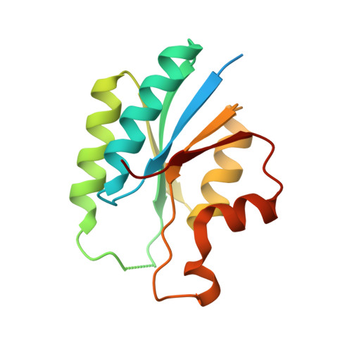
7JI4 - PubMed Abstract:
The KdpDE two-component system regulates potassium homeostasis and virulence in various bacterial species. The KdpD histidine kinases (HK) of this system contain a universal stress protein (USP) domain which binds to the second messenger cyclic-di-adenosine monophosphate (c-di-AMP) for regulating transcriptional output from this two-component system in Firmicutes such as Staphylococcus aureus. However, the structural basis of c-di-AMP specificity within the KdpD-USP domain is not well understood. Here, we resolved a 2.3 Å crystal structure of the S. aureus KdpD-USP domain (USP Sa ) complexed with c-di-AMP. Binding affinity analyses of USP Sa mutants targeting the observed USP Sa :c-di-AMP structural interface enabled the identification of the sequence residues that are required for c-di-AMP specificity. Based on the conservation of these residues in other Firmicutes, we identified the binding motif, (A/G/C)XSXSX 2 N(Y/F), which allowed us to predict c-di-AMP binding in other KdpD HKs. Furthermore, we found that the USP Sa domain contains structural features distinct from the canonical standalone USPs that bind ATP as a preferred ligand. These features include inward-facing conformations of its β1-α1 and β4-α4 loops, a short α2 helix, the absence of a triphosphate-binding Walker A motif, and a unique dual phospho-ligand binding mode. It is therefore likely that USP Sa -like domains in KdpD HKs represent a novel subfamily of the USPs.
- Department of Medical and Molecular Sciences, University of Delaware, Newark, Delaware, USA.
Organizational Affiliation: