7FH3 | pdb_00007fh3

Crystal structure of the ATP sulfurylase domain of human PAPSS2


Experimental Data Snapshot

  • Method: X-RAY DIFFRACTION
  • Resolution: 1.80 Å
  • R-Value Free: 
    0.199 (Depositor), 0.201 (DCC) 
  • R-Value Work: 
    0.169 (Depositor), 0.172 (DCC) 
  • R-Value Observed: 
    0.170 (Depositor) 

Starting Model: experimental
View more details

wwPDB Validation 3D Report Full Report

Validation slider image for 7FH3

Ligand Structure Quality Assessment 


This is version 1.2 of the entry. See complete history

Literature

Structural basis for the substrate recognition mechanism of ATP-sulfurylase domain of human PAPS synthase 2.

Zhang, P.Zhang, L.Hou, Z.Lin, H.Gao, H.Zhang, L.

(2022) Biochem Biophys Res Commun 586: 1-7

  • DOI: https://doi.org/10.1016/j.bbrc.2021.11.062
  • Primary Citation Related Structures: 
    7FH3, 7FHA

  • PubMed Abstract: 

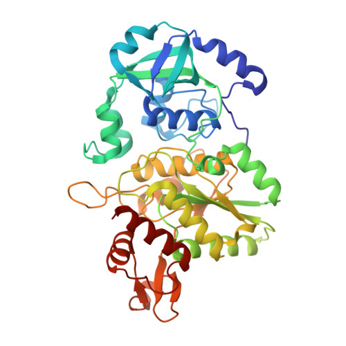
    Sulfation is an essential modification on biomolecules in living cells, and 3'-Phosphoadenosine-5'-phosphosulfate (PAPS) is its unique and universal sulfate donor. Human PAPS synthases (PAPSS1 and 2) are the only enzymes that catalyze PAPS production from inorganic sulfate. Unexpectedly, PAPSS1 and PAPSS2 do not functional complement with each other, and abnormal function of PAPSS2 but not PAPSS1 leads to numerous human diseases including bone development diseases, hormone disorder and cancers. Here, we reported the crystal structures of ATP-sulfurylase domain of human PAPSS2 (ATPS2) and ATPS2 in complex with is product 5'-phosphosulfate (APS). We demonstrated that ATPS2 recognizes the substrates by using family conserved residues located on the HXXH and PP motifs, and achieves substrate binding and releasing by employing a non-conserved phenylalanine (Phe550) through a never observed flipping mechanism. Our discovery provides additional information to better understand the biological function of PAPSS2 especially in tumorigenesis, and may facilitate the drug discovery against this enzyme.


  • Organizational Affiliation
    • Key Laboratory of Medical Epigenetics and Metabolism, Institutes of Biomedical Sciences, Shanghai Medical College, Fudan University, Shanghai, 200032, China.

Macromolecule Content 

  • Total Structure Weight: 91.55 kDa 
  • Atom Count: 7,237 
  • Modeled Residue Count: 782 
  • Deposited Residue Count: 786 
  • Unique protein chains: 1

Macromolecules

Find similar proteins by:|  3D Structure
Entity ID: 1
MoleculeChains  Sequence LengthOrganismDetailsImage
Bifunctional 3'-phosphoadenosine 5'-phosphosulfate synthase 2
A, B
393Homo sapiensMutation(s): 0 
Gene Names: PAPSS2ATPSK2
EC: 2.7.7.4 (PDB Primary Data), 2.7.1.25 (PDB Primary Data)
UniProt & NIH Common Fund Data Resources
Find proteins for O95340 (Homo sapiens)
Explore O95340 
Go to UniProtKB:  O95340
PHAROS:  O95340
GTEx:  ENSG00000198682 
Entity Groups
Sequence Clusters30% Identity50% Identity70% Identity90% Identity95% Identity100% Identity
UniProt GroupO95340
Sequence Annotations
Expand
Reference Sequence

Small Molecules

Ligands 2 Unique
IDChains Name / Formula / InChI Key2D Diagram3D Interactions
BGC
(Subject of Investigation/LOI)

Query on BGC


Download:Ideal Coordinates CCD File
J [auth A],
P [auth B]
beta-D-glucopyranose
C6 H12 O6
WQZGKKKJIJFFOK-VFUOTHLCSA-N
SO4
(Subject of Investigation/LOI)

Query on SO4


Download:Ideal Coordinates CCD File
C [auth A]
D [auth A]
E [auth A]
F [auth A]
G [auth A]
C [auth A],
D [auth A],
E [auth A],
F [auth A],
G [auth A],
H [auth A],
I [auth A],
K [auth B],
L [auth B],
M [auth B],
N [auth B],
O [auth B]
SULFATE ION
O4 S
QAOWNCQODCNURD-UHFFFAOYSA-L

Experimental Data & Validation

Experimental Data

  • Method: X-RAY DIFFRACTION
  • Resolution: 1.80 Å
  • R-Value Free:  0.199 (Depositor), 0.201 (DCC) 
  • R-Value Work:  0.169 (Depositor), 0.172 (DCC) 
  • R-Value Observed: 0.170 (Depositor) 
Space Group: H 3
Unit Cell:
Length ( Å )Angle ( ˚ )
a = 179.859α = 90
b = 179.859β = 90
c = 79.289γ = 120
Software Package:
Software NamePurpose
PHENIXrefinement
PDB_EXTRACTdata extraction
HKL-3000data reduction
HKL-3000data scaling
PHASERphasing

Structure Validation

View Full Validation Report


Ligand Structure Quality Assessment 


Entry History 

& Funding Information

Deposition Data


Funding OrganizationLocationGrant Number
National Natural Science Foundation of China (NSFC)China91853118
National Natural Science Foundation of China (NSFC)China22077081
National Natural Science Foundation of China (NSFC)China21722802

Revision History  (Full details and data files)

  • Version 1.0: 2021-12-01
    Type: Initial release
  • Version 1.1: 2022-02-16
    Changes: Database references
  • Version 1.2: 2023-11-29
    Changes: Data collection, Refinement description