7F7Y

Crystal Structure of protein PitB from pilus islet-2 of Streptococcus pneumoniae


Experimental Data Snapshot

  • Method: X-RAY DIFFRACTION
  • Resolution: 2.13 Å
  • R-Value Free: 0.235 
  • R-Value Work: 0.198 
  • R-Value Observed: 0.200 

wwPDB Validation   3D Report Full Report


Ligand Structure Quality Assessment 


This is version 1.3 of the entry. See complete history


Literature

New structural insights into the PI-2 pilus from Streptococcus oralis, an early dental plaque colonizer.

Yadav, R.K.Krishnan, V.

(2022) FEBS J 289: 6342-6366

  • DOI: https://doi.org/10.1111/febs.16527
  • Primary Citation of Related Structures:  
    7F7Y, 7VCN, 7VCR, 7W6B, 7W7I

  • PubMed Abstract: 

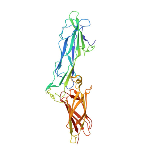
    Streptococcus oralis is a member of the mitis group of oral streptococci and an early colonizer in dental plaque biofilm, a major cause of periodontal disease, dental caries, and other oral infections. S. oralis promotes biofilm growth by coaggregating in a mutualistic partnership with other early colonizers such as Actinomyces oris. For this cell-to-cell interaction, A. oris is known to use its sortase-dependent pilus (type 2), but whether S. oralis uses its PI-2 (pilus islet 2) pilus is still to be determined. The PI-2 pilus is predicted to have a heterodimeric structure consisting of two different protein subunits with their own location and function: the tip PitA pilin for adhesion and the backbone PitB pilin for length. Thus far, structural information remains incomplete about the role of PI-2 pili in the mutualistic mechanism between S. oralis and A. oris. We now report on the crystal structure analysis of PitA and PitB using X-ray crystallography, small-angle X-ray scattering, and molecular docking studies. Accordingly, we propose a structural model for the PI-2 pilus, wherein repeating PitB subunits are arranged head-to-tail to form the long backbone structure with PitA on the outer tip. By performing both in vitro and in vivo experiments, we examined the role played by PitA in mediating the mutualistic interaction between S. oralis and A. oris, which appears to involve the coaggregation factor CafA. We also reveal that the galactose monosaccharide is a conceivable ligand for PitA and thereby might be used to inhibit coaggregation and control oral biofilm development. DATABASE: Structural coordinates for the PitA fragment, PitA fragment TbXO4 derivative, full-length PitA, and PitB from S. oralis have been deposited at the Protein Data Bank as 7VCR, 7W7I, 7VCN, 7W6B, and 7W7I, respectively. Streptococcus pneumoniae PitB coordinates have been deposited as 7F7Y.


  • Organizational Affiliation

    Laboratory of Structural Microbiology, Regional Centre for Biotechnology, NCR Biotech Science Cluster, Faridabad, India.


Macromolecules
Find similar proteins by:  (by identity cutoff)  |  3D Structure
Entity ID: 1
MoleculeChains Sequence LengthOrganismDetailsImage
PitB
A, B
332Streptococcus pneumoniaeMutation(s): 0 
Gene Names: pitB
UniProt
Find proteins for B3FNT1 (Streptococcus pneumoniae)
Explore B3FNT1 
Go to UniProtKB:  B3FNT1
Entity Groups  
Sequence Clusters30% Identity50% Identity70% Identity90% Identity95% Identity100% Identity
UniProt GroupB3FNT1
Sequence Annotations
Expand
  • Reference Sequence
Small Molecules
Ligands 3 Unique
IDChains Name / Formula / InChI Key2D Diagram3D Interactions
IOD (Subject of Investigation/LOI)
Query on IOD

Download Ideal Coordinates CCD File 
C [auth A]
F [auth A]
G [auth A]
H [auth A]
I [auth A]
C [auth A],
F [auth A],
G [auth A],
H [auth A],
I [auth A],
J [auth A],
K [auth B],
O [auth B],
P [auth B]
IODIDE ION
I
XMBWDFGMSWQBCA-UHFFFAOYSA-M
SO4 (Subject of Investigation/LOI)
Query on SO4

Download Ideal Coordinates CCD File 
D [auth A],
E [auth A]
SULFATE ION
O4 S
QAOWNCQODCNURD-UHFFFAOYSA-L
K (Subject of Investigation/LOI)
Query on K

Download Ideal Coordinates CCD File 
L [auth B],
M [auth B],
N [auth B]
POTASSIUM ION
K
NPYPAHLBTDXSSS-UHFFFAOYSA-N
Experimental Data & Validation

Experimental Data

  • Method: X-RAY DIFFRACTION
  • Resolution: 2.13 Å
  • R-Value Free: 0.235 
  • R-Value Work: 0.198 
  • R-Value Observed: 0.200 
  • Space Group: C 2 2 21
Unit Cell:
Length ( Å )Angle ( ˚ )
a = 73.49α = 90
b = 82.659β = 90
c = 362.193γ = 90
Software Package:
Software NamePurpose
REFMACrefinement
MOSFLMdata reduction
SCALAdata scaling
MOLREPphasing

Structure Validation

View Full Validation Report


Ligand Structure Quality Assessment 


Entry History & Funding Information

Deposition Data


Funding OrganizationLocationGrant Number
Science and Engineering Research Board (SERB)IndiaCRG/2019/000432

Revision History  (Full details and data files)

  • Version 1.0: 2022-06-01
    Type: Initial release
  • Version 1.1: 2022-06-08
    Changes: Database references
  • Version 1.2: 2022-10-26
    Changes: Database references, Derived calculations
  • Version 1.3: 2023-11-29
    Changes: Data collection, Refinement description