7F5R | pdb_00007f5r

Crystal structure of SARS-CoV-2 Y453F-RBD bound to mink ACE2


Experimental Data Snapshot

  • Method: X-RAY DIFFRACTION
  • Resolution: 3.01 Å
  • R-Value Free: 
    0.218 (Depositor), 0.220 (DCC) 
  • R-Value Work: 
    0.181 (Depositor), 0.180 (DCC) 
  • R-Value Observed: 
    0.183 (Depositor) 

Starting Model: experimental
View more details

wwPDB Validation   3D Report Full Report


Ligand Structure Quality Assessment 


This is version 1.3 of the entry. See complete history


Literature

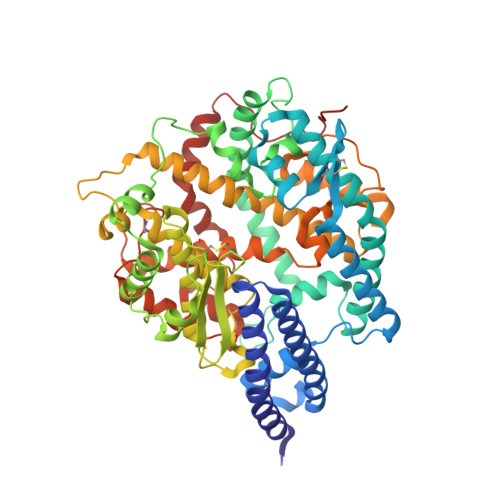
Mutation Y453F in the spike protein of SARS-CoV-2 enhances interaction with the mink ACE2 receptor for host adaption.

Ren, W.Lan, J.Ju, X.Gong, M.Long, Q.Zhu, Z.Yu, Y.Wu, J.Zhong, J.Zhang, R.Fan, S.Zhong, G.Huang, A.Wang, X.Ding, Q.

(2021) PLoS Pathog 17: e1010053-e1010053

  • DOI: https://doi.org/10.1371/journal.ppat.1010053
  • Primary Citation of Related Structures:  
    7F5R

  • PubMed Abstract: 

    COVID-19 patients transmitted SARS-CoV-2 to minks in the Netherlands in April 2020. Subsequently, the mink-associated virus (miSARS-CoV-2) spilled back over into humans. Genetic sequences of the miSARS-CoV-2 identified a new genetic variant known as "Cluster 5" that contained mutations in the spike protein. However, the functional properties of these "Cluster 5" mutations have not been well established. In this study, we found that the Y453F mutation located in the RBD domain of miSARS-CoV-2 is an adaptive mutation that enhances binding to mink ACE2 and other orthologs of Mustela species without compromising, and even enhancing, its ability to utilize human ACE2 as a receptor for entry. Structural analysis suggested that despite the similarity in the overall binding mode of SARS-CoV-2 RBD to human and mink ACE2, Y34 of mink ACE2 was better suited to interact with a Phe rather than a Tyr at position 453 of the viral RBD due to less steric clash and tighter hydrophobic-driven interaction. Additionally, the Y453F spike exhibited resistance to convalescent serum, posing a risk for vaccine development. Thus, our study suggests that since the initial transmission from humans, SARS-CoV-2 evolved to adapt to the mink host, leading to widespread circulation among minks while still retaining its ability to efficiently utilize human ACE2 for entry, thus allowing for transmission of the miSARS-CoV-2 back into humans. These findings underscore the importance of active surveillance of SARS-CoV-2 evolution in Mustela species and other susceptible hosts in order to prevent future outbreaks.


  • Organizational Affiliation
    • Center for Infectious Disease Research, School of Medicine, Tsinghua University, Beijing, China.

Macromolecules
Find similar proteins by:  (by identity cutoff)  |  3D Structure
Entity ID: 1
MoleculeChains Sequence LengthOrganismDetailsImage
mink ACE2A,
C [auth B],
E [auth D]
597Neogale visonMutation(s): 0 
EC: 3.4
UniProt
Find proteins for A0A8C7BTF2 (Neovison vison)
Explore A0A8C7BTF2 
Go to UniProtKB:  A0A8C7BTF2
Entity Groups  
Sequence Clusters30% Identity50% Identity70% Identity90% Identity95% Identity100% Identity
UniProt GroupA0A8C7BTF2
Glycosylation
Glycosylation Sites: 2
Sequence Annotations
Expand
  • Reference Sequence
Find similar proteins by:  (by identity cutoff)  |  3D Structure
Entity ID: 2
MoleculeChains Sequence LengthOrganismDetailsImage
Spike protein S1B [auth E],
D [auth C],
F
197Severe acute respiratory syndrome coronavirus 2Mutation(s): 1 
Gene Names: S2
UniProt
Find proteins for P0DTC2 (Severe acute respiratory syndrome coronavirus 2)
Explore P0DTC2 
Go to UniProtKB:  P0DTC2
Entity Groups  
Sequence Clusters30% Identity50% Identity70% Identity90% Identity95% Identity100% Identity
UniProt GroupP0DTC2
Glycosylation
Glycosylation Sites: 1Go to GlyGen: P0DTC2-1
Sequence Annotations
Expand
  • Reference Sequence
Oligosaccharides

Help

Entity ID: 3
MoleculeChains Length2D Diagram Glycosylation3D Interactions
2-acetamido-2-deoxy-beta-D-glucopyranose-(1-4)-2-acetamido-2-deoxy-beta-D-glucopyranose
G, I
2N-Glycosylation
Glycosylation Resources
GlyTouCan:  G42666HT
GlyCosmos:  G42666HT
GlyGen:  G42666HT
Entity ID: 4
MoleculeChains Length2D Diagram Glycosylation3D Interactions
alpha-L-fucopyranose-(1-6)-2-acetamido-2-deoxy-beta-D-glucopyranose
H
2N-Glycosylation
Glycosylation Resources
GlyTouCan:  G86851RC
GlyCosmos:  G86851RC
GlyGen:  G86851RC
Experimental Data & Validation

Experimental Data

  • Method: X-RAY DIFFRACTION
  • Resolution: 3.01 Å
  • R-Value Free:  0.218 (Depositor), 0.220 (DCC) 
  • R-Value Work:  0.181 (Depositor), 0.180 (DCC) 
  • R-Value Observed: 0.183 (Depositor) 
Space Group: P 21 21 2
Unit Cell:
Length ( Å )Angle ( ˚ )
a = 290.185α = 90
b = 130.185β = 90
c = 136.887γ = 90
Software Package:
Software NamePurpose
PHENIXrefinement
PDB_EXTRACTdata extraction
HKL-2000data reduction
HKL-2000data scaling
MOLREPphasing

Structure Validation

View Full Validation Report


Ligand Structure Quality Assessment 


Entry History & Funding Information

Deposition Data


Funding OrganizationLocationGrant Number
Not funded--

Revision History  (Full details and data files)

  • Version 1.0: 2022-06-22
    Type: Initial release
  • Version 1.1: 2023-11-29
    Changes: Data collection, Refinement description
  • Version 1.2: 2024-10-09
    Changes: Structure summary
  • Version 1.3: 2025-06-18
    Changes: Database references