7F5M

Crystal structure of Sas5 YEATS domain in complex with H3K27bz peptide


Experimental Data Snapshot

  • Method: X-RAY DIFFRACTION
  • Resolution: 2.40 Å
  • R-Value Free: 0.274 
  • R-Value Work: 0.227 
  • R-Value Observed: 0.229 

wwPDB Validation   3D Report Full Report


This is version 1.2 of the entry. See complete history


Literature

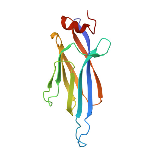
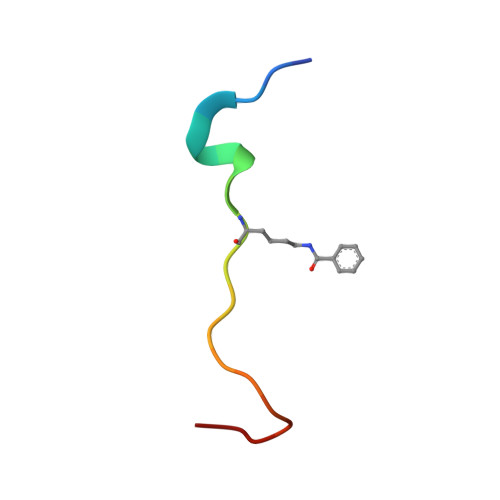
Global profiling of regulatory elements in the histone benzoylation pathway.

Wang, D.Yan, F.Wu, P.Ge, K.Li, M.Li, T.Gao, Y.Peng, C.Chen, Y.

(2022) Nat Commun 13: 1369-1369

  • DOI: https://doi.org/10.1038/s41467-022-29057-2
  • Primary Citation of Related Structures:  
    7F3S, 7F4A, 7F4E, 7F51, 7F5M

  • PubMed Abstract: 

    Lysine benzoylation (Kbz) is a recently discovered post-translational modification associated with active transcription. However, the proteins for maintaining and interpreting Kbz and the physiological roles of Kbz remain elusive. Here, we systematically characterize writer, eraser, and reader proteins of histone Kbz in S. cerevisiae using proteomic, biochemical, and structural approaches. Our study identifies 27 Kbz sites on yeast histones that can be regulated by cellular metabolic states. The Spt-Ada-Gcn5 acetyltransferase (SAGA) complex and NAD + -dependent histone deacetylase Hst2 could function as the writer and eraser of histone Kbz, respectively. Crystal structures of Hst2 complexes reveal the molecular basis for Kbz recognition and catalysis by Hst2. In addition, we demonstrate that a subset of YEATS domains and bromodomains serve as Kbz readers, and structural analyses reveal how YEATS and bromodomains recognize Kbz marks. Moreover, the proteome-wide screening of Kbz-modified proteins identifies 207 Kbz sites on 149 non-histone proteins enriched in ribosome biogenesis, glycolysis/gluconeogenesis, and rRNA processing pathways. Our studies identify regulatory elements for the Kbz pathway and provide a framework for dissecting the biological functions of lysine benzoylation.


  • Organizational Affiliation

    State Key Laboratory of Molecular Biology, Shanghai Institute of Biochemistry and Cell Biology, Center for Excellence in Molecular Cell Science, Chinese Academy of Sciences, 200031, Shanghai, China.


Macromolecules
Find similar proteins by:  (by identity cutoff)  |  3D Structure
Entity ID: 1
MoleculeChains Sequence LengthOrganismDetailsImage
Something about silencing protein 5
A, B
139Saccharomyces cerevisiae S288CMutation(s): 0 
Gene Names: SAS5YOR213C
UniProt
Find proteins for Q99314 (Saccharomyces cerevisiae (strain ATCC 204508 / S288c))
Explore Q99314 
Go to UniProtKB:  Q99314
Entity Groups  
Sequence Clusters30% Identity50% Identity70% Identity90% Identity95% Identity100% Identity
UniProt GroupQ99314
Sequence Annotations
Expand
  • Reference Sequence

Find similar proteins by:  Sequence   |   3D Structure  

Entity ID: 2
MoleculeChains Sequence LengthOrganismDetailsImage
LYS-GLN-LEU-ALA-SER-LYS-ALA-ALA-ARG-LBZ-SER-ALA-PRO-SER-THR-GLY-GLY-VAL-LYS-TYR20Saccharomyces cerevisiaeMutation(s): 0 
UniProt
Find proteins for P61830 (Saccharomyces cerevisiae (strain ATCC 204508 / S288c))
Explore P61830 
Go to UniProtKB:  P61830
Entity Groups  
Sequence Clusters30% Identity50% Identity70% Identity90% Identity95% Identity100% Identity
UniProt GroupP61830
Sequence Annotations
Expand
  • Reference Sequence
Experimental Data & Validation

Experimental Data

  • Method: X-RAY DIFFRACTION
  • Resolution: 2.40 Å
  • R-Value Free: 0.274 
  • R-Value Work: 0.227 
  • R-Value Observed: 0.229 
  • Space Group: P 32 2 1
Unit Cell:
Length ( Å )Angle ( ˚ )
a = 126.578α = 90
b = 126.578β = 90
c = 48.708γ = 120
Software Package:
Software NamePurpose
PHENIXrefinement
HKL-2000data scaling
PDB_EXTRACTdata extraction
HKL-2000data reduction
PHASESphasing

Structure Validation

View Full Validation Report


Entry History & Funding Information

Deposition Data


Funding OrganizationLocationGrant Number
Chinese Academy of SciencesChinaXDB37010303
National Natural Science Foundation of China (NSFC)China31970576

Revision History  (Full details and data files)

  • Version 1.0: 2022-03-16
    Type: Initial release
  • Version 1.1: 2022-03-30
    Changes: Database references
  • Version 1.2: 2023-11-29
    Changes: Data collection, Derived calculations, Refinement description