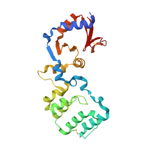
Crystal structure of the antibiotic- and nitrite-responsive histidine kinase VbrK sensor domain from Vibrio rotiferianus.
Cho, S.Y., Yoon, S.I.(2021) Biochem Biophys Res Commun 568: 136-142
- PubMed: 34214877
- DOI: https://doi.org/10.1016/j.bbrc.2021.06.076
- Primary Citation of Related Structures:
7F2G, 7F2H - PubMed Abstract:
Vibrio species are prevalent in the aquatic environments and can infect humans and aquatic organisms. Vibrio parahaemolyticus counteracts β-lactam antibiotics and enhances virulence using a regulation mechanism mediated by a two-component regulatory system (TCS) consisting of the VbrK histidine kinase and the VbrR response regulator. The periplasmic sensor domain of VbrK (VbrK SD ) detects β-lactam antibiotics or undergoes S-nitrosylation in response to host nitrites. Although V. parahaemolyticus VbrK SD (vpVbrK SD ) has recently been characterized through structural studies, it is unclear whether its structural features that are indispensable for biological functions are conserved in other VbrK orthologs. To structurally define the functionally critical regions of VbrK and address the structural dynamics of VbrK, we determined the crystal structures of Vibrio rotiferianus VbrK SD (vrVbrK SD ) in two crystal forms and performed a comparative analysis of diverse VbrK structures. vrVbrK SD folds into a curved rod-shaped two-domain structure as observed in vpVbrK SD . The membrane-distal end of the vrVbrK SD structure, including the α3 helix and its neighboring loops, harbors both S-nitrosylation and antibiotic-sensing sites and displays high structural flexibility and diversity. Noticeably, the distal end is partially stabilized by a disulfide bond, which is formed by the cysteine residue that is S-nitrosylated in response to nitrite. Therefore, the distal end of VbrK SD plays a key role in initiating the VbrK-VbrR TCS pathway activation, and it is involved in the nitrosylation-mediated regulation of the structural dynamics of VbrK.
- Division of Biomedical Convergence, College of Biomedical Science, Kangwon National University, Chuncheon, 24341, Republic of Korea.
Organizational Affiliation:


