7EPT

Structural basis for the tethered peptide activation of adhesion GPCRs


Experimental Data Snapshot

  • Method: ELECTRON MICROSCOPY
  • Resolution: 3.00 Å
  • Aggregation State: PARTICLE 
  • Reconstruction Method: SINGLE PARTICLE 

wwPDB Validation   3D Report Full Report


This is version 1.0 of the entry. See complete history


Literature

Structural basis for the tethered peptide activation of adhesion GPCRs.

Ping, Y.Q.Xiao, P.Yang, F.Zhao, R.J.Guo, S.C.Yan, X.Wu, X.Zhang, C.Lu, Y.Zhao, F.Zhou, F.Xi, Y.T.Yin, W.Liu, F.Z.He, D.F.Zhang, D.L.Zhu, Z.L.Jiang, Y.Du, L.Feng, S.Q.Schoneberg, T.Liebscher, I.Xu, H.E.Sun, J.P.

(2022) Nature 604: 763-770

  • DOI: https://doi.org/10.1038/s41586-022-04619-y
  • Primary Citation of Related Structures:  
    7EPT, 7EQ1

  • PubMed Abstract: 

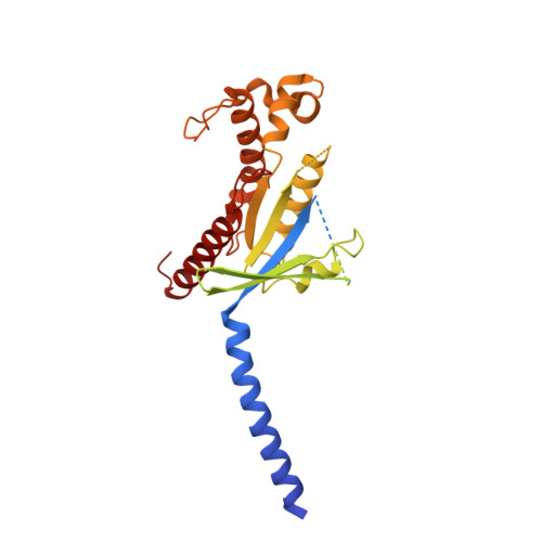
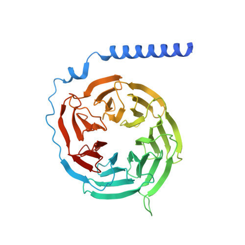
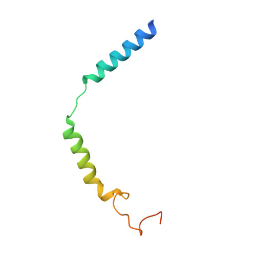
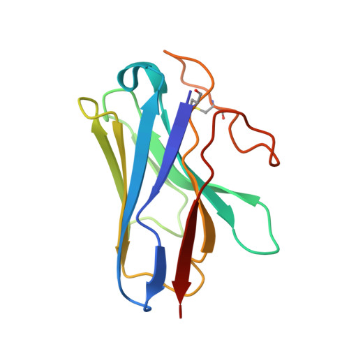
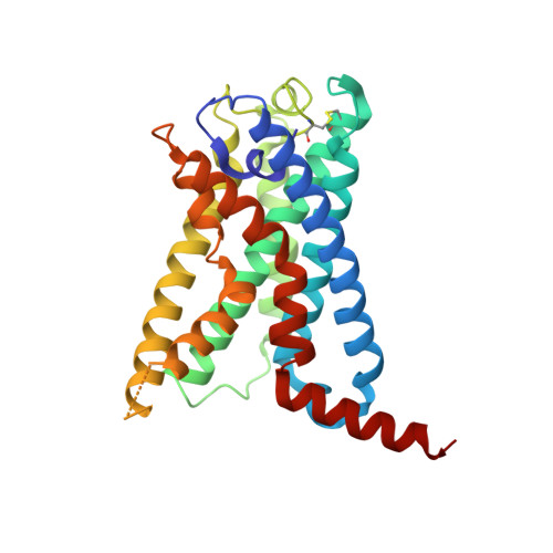
    Adhesion G-protein-coupled receptors (aGPCRs) are important for organogenesis, neurodevelopment, reproduction and other processes 1-6 . Many aGPCRs are activated by a conserved internal (tethered) agonist sequence known as the Stachel sequence 7-12 . Here, we report the cryogenic electron microscopy (cryo-EM) structures of two aGPCRs in complex with G s : GPR133 and GPR114. The structures indicate that the Stachel sequences of both receptors assume an α-helical-bulge-β-sheet structure and insert into a binding site formed by the transmembrane domain (TMD). A hydrophobic interaction motif (HIM) within the Stachel sequence mediates most of the intramolecular interactions with the TMD. Combined with the cryo-EM structures, biochemical characterization of the HIM motif provides insight into the cross-reactivity and selectivity of the Stachel sequences. Two interconnected mechanisms, the sensing of Stachel sequences by the conserved 'toggle switch' W 6.53 and the constitution of a hydrogen-bond network formed by Q 7.49 /Y 7.49 and the P 6.47 /V 6.47 φφG 6.50 motif (φ indicates a hydrophobic residue), are important in Stachel sequence-mediated receptor activation and G s coupling. Notably, this network stabilizes kink formation in TM helices 6 and 7 (TM6 and TM7, respectively). A common G s -binding interface is observed between the two aGPCRs, and GPR114 has an extended TM7 that forms unique interactions with G s . Our structures reveal the detailed mechanisms of aGPCR activation by Stachel sequences and their G s coupling.


  • Organizational Affiliation

    Key Laboratory Experimental Teratology of the Ministry of Education, Department of Biochemistry and Molecular Biology, School of Basic Medical Sciences, Cheeloo College of Medicine, Shandong University, Jinan, China.


Macromolecules
Find similar proteins by:  (by identity cutoff)  |  3D Structure
Entity ID: 1
MoleculeChains Sequence LengthOrganismDetailsImage
Guanine nucleotide-binding protein G(s) subunit alpha isoforms short394Homo sapiensMutation(s): 0 
Gene Names: GPR133ADGRD1PGR25
Membrane Entity: Yes 
UniProt & NIH Common Fund Data Resources
Find proteins for P63092 (Homo sapiens)
Explore P63092 
Go to UniProtKB:  P63092
PHAROS:  P63092
GTEx:  ENSG00000087460 
Entity Groups  
Sequence Clusters30% Identity50% Identity70% Identity90% Identity95% Identity100% Identity
UniProt GroupP63092
Sequence Annotations
Expand
  • Reference Sequence
Find similar proteins by:  (by identity cutoff)  |  3D Structure
Entity ID: 2
MoleculeChains Sequence LengthOrganismDetailsImage
Guanine nucleotide-binding protein G(I)/G(S)/G(T) subunit beta-1358Homo sapiensMutation(s): 0 
Gene Names: GNB1
UniProt & NIH Common Fund Data Resources
Find proteins for P62873 (Homo sapiens)
Explore P62873 
Go to UniProtKB:  P62873
PHAROS:  P62873
GTEx:  ENSG00000078369 
Entity Groups  
Sequence Clusters30% Identity50% Identity70% Identity90% Identity95% Identity100% Identity
UniProt GroupP62873
Sequence Annotations
Expand
  • Reference Sequence
Find similar proteins by:  (by identity cutoff)  |  3D Structure
Entity ID: 3
MoleculeChains Sequence LengthOrganismDetailsImage
Guanine nucleotide-binding protein G(I)/G(S)/G(O) subunit gamma-2C [auth G]71Homo sapiensMutation(s): 0 
Gene Names: GNG2
Membrane Entity: Yes 
UniProt & NIH Common Fund Data Resources
Find proteins for P59768 (Homo sapiens)
Explore P59768 
Go to UniProtKB:  P59768
PHAROS:  P59768
GTEx:  ENSG00000186469 
Entity Groups  
Sequence Clusters30% Identity50% Identity70% Identity90% Identity95% Identity100% Identity
UniProt GroupP59768
Sequence Annotations
Expand
  • Reference Sequence
Find similar proteins by:  (by identity cutoff)  |  3D Structure
Entity ID: 4
MoleculeChains Sequence LengthOrganismDetailsImage
Nanobody-35D [auth N]128synthetic constructMutation(s): 0 
Entity Groups  
Sequence Clusters30% Identity50% Identity70% Identity90% Identity95% Identity100% Identity
Sequence Annotations
Expand
  • Reference Sequence
Find similar proteins by:  (by identity cutoff)  |  3D Structure
Entity ID: 5
MoleculeChains Sequence LengthOrganismDetailsImage
Adhesion G-protein coupled receptor D1E [auth R]283Homo sapiensMutation(s): 0 
Gene Names: ADGRD1GPR133PGR25
Membrane Entity: Yes 
UniProt & NIH Common Fund Data Resources
Find proteins for Q6QNK2 (Homo sapiens)
Explore Q6QNK2 
Go to UniProtKB:  Q6QNK2
PHAROS:  Q6QNK2
GTEx:  ENSG00000111452 
Entity Groups  
Sequence Clusters30% Identity50% Identity70% Identity90% Identity95% Identity100% Identity
UniProt GroupQ6QNK2
Sequence Annotations
Expand
  • Reference Sequence
Small Molecules
Ligands 1 Unique
IDChains Name / Formula / InChI Key2D Diagram3D Interactions
PLM
Query on PLM

Download Ideal Coordinates CCD File 
F [auth R]PALMITIC ACID
C16 H32 O2
IPCSVZSSVZVIGE-UHFFFAOYSA-N
Experimental Data & Validation

Experimental Data

  • Method: ELECTRON MICROSCOPY
  • Resolution: 3.00 Å
  • Aggregation State: PARTICLE 
  • Reconstruction Method: SINGLE PARTICLE 

Structure Validation

View Full Validation Report


Entry History & Funding Information

Deposition Data


Funding OrganizationLocationGrant Number
National Natural Science Foundation of China (NSFC)China--

Revision History  (Full details and data files)

  • Version 1.0: 2022-05-11
    Type: Initial release