7EPQ

Crystal structure of exopolyphosphatase (PPX) from Porphyromonas gingivalis in complex with sulfate and magnesium ions


Experimental Data Snapshot

  • Method: X-RAY DIFFRACTION
  • Resolution: 2.20 Å
  • R-Value Free: 0.240 
  • R-Value Work: 0.180 

wwPDB Validation   3D Report Full Report


Ligand Structure Quality Assessment 


This is version 1.1 of the entry. See complete history


Literature

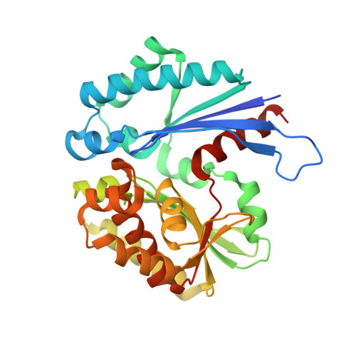
The structure of exopolyphosphatase (PPX) from Porphyromonas gingivalis in complex with substrate analogs and magnesium ions reveals the basis for polyphosphate processivity.

Zhang, A.Lu, Z.Xu, Y.Qi, T.Li, W.Zhang, L.Cui, Z.

(2021) J Struct Biol 213: 107767-107767

  • DOI: https://doi.org/10.1016/j.jsb.2021.107767
  • Primary Citation of Related Structures:  
    7EPQ

  • PubMed Abstract: 

    The enzymes exopolyphosphatase/guanosine pentaphosphate phosphohydrolase (PPX/GppA) play important roles in the bacterial stringent response. PPX degrades inorganic polyphosphate (polyP), a polymer composed of a few to hundreds of phosphate residues supporting cell survival in the stationary phase. The crystal structure of PPX from Porphyromonas gingivalis (PgPPX) in complex with catalytic magnesium ions and several sulfate ions was solved. PgPPX contained two domains and represented a "closed" configuration. Four sulfate ions forming a linear dispersed chain were observed in the aqueduct of the PPX dimer, which the long polyP chain most likely occupied. The side chain of R255 stretched into the cavity where polyP could be located, obstructing the entrance of larger substrates such as NTP and NDP. This study provided the first view into the structure of the PPX/GppA homolog in complex with magnesium ions and substrate analogs and explained how PgPPX implemented its functionality.


  • Organizational Affiliation

    Food and Pharmacy College, Xuchang University, China; Key Laboratory of Biomarker Based Rapid-detection Technology for Food Safety of Henan Province, Xuchang University, China.


Macromolecules
Find similar proteins by:  (by identity cutoff)  |  3D Structure
Entity ID: 1
MoleculeChains Sequence LengthOrganismDetailsImage
Putative exopolyphosphatase
A, B
307Porphyromonas gingivalis ATCC 33277Mutation(s): 0 
Gene Names: PGN_0378
UniProt
Find proteins for B2RHQ2 (Porphyromonas gingivalis (strain ATCC 33277 / DSM 20709 / CIP 103683 / JCM 12257 / NCTC 11834 / 2561))
Explore B2RHQ2 
Go to UniProtKB:  B2RHQ2
Entity Groups  
Sequence Clusters30% Identity50% Identity70% Identity90% Identity95% Identity100% Identity
UniProt GroupB2RHQ2
Sequence Annotations
Expand
  • Reference Sequence
Small Molecules
Ligands 2 Unique
IDChains Name / Formula / InChI Key2D Diagram3D Interactions
SO4 (Subject of Investigation/LOI)
Query on SO4

Download Ideal Coordinates CCD File 
C [auth A]
D [auth A]
E [auth A]
F [auth A]
J [auth B]
C [auth A],
D [auth A],
E [auth A],
F [auth A],
J [auth B],
K [auth B],
L [auth B],
M [auth B]
SULFATE ION
O4 S
QAOWNCQODCNURD-UHFFFAOYSA-L
MG (Subject of Investigation/LOI)
Query on MG

Download Ideal Coordinates CCD File 
G [auth A]
H [auth A]
I [auth A]
N [auth B]
O [auth B]
G [auth A],
H [auth A],
I [auth A],
N [auth B],
O [auth B],
P [auth B],
Q [auth B]
MAGNESIUM ION
Mg
JLVVSXFLKOJNIY-UHFFFAOYSA-N
Experimental Data & Validation

Experimental Data

  • Method: X-RAY DIFFRACTION
  • Resolution: 2.20 Å
  • R-Value Free: 0.240 
  • R-Value Work: 0.180 
  • Space Group: P 21 21 21
Unit Cell:
Length ( Å )Angle ( ˚ )
a = 64.43α = 90
b = 86.61β = 90
c = 122.92γ = 90
Software Package:
Software NamePurpose
HKL-2000data scaling
PHENIXrefinement
PDB_EXTRACTdata extraction
HKL-2000data reduction
BALBESphasing

Structure Validation

View Full Validation Report


Ligand Structure Quality Assessment 


Entry History 

Deposition Data

  • Released Date: 2022-05-11 
  • Deposition Author(s): Zhang, A.

Revision History  (Full details and data files)

  • Version 1.0: 2022-05-11
    Type: Initial release
  • Version 1.1: 2023-11-29
    Changes: Data collection, Refinement description