Crystal structure of an apo 7 alpha-hydroxysteroid dehydrogenase reveals key structural changes induced by substrate and co-factor binding.
Kim, K.H., Lee, C.W., Pardhe, B.D., Hwang, J., Do, H., Lee, Y.M., Lee, J.H., Oh, T.J.(2021) J Steroid Biochem Mol Biol 212: 105945-105945
- PubMed: 34171491 Search on PubMed
- DOI: https://doi.org/10.1016/j.jsbmb.2021.105945
- Primary Citation Related Structures:
7ENY - PubMed Abstract:
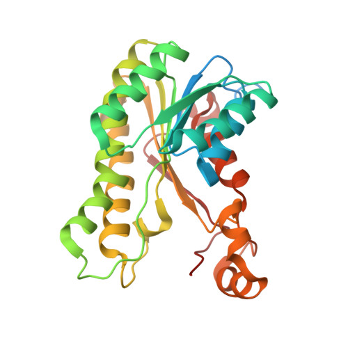
7α-Hydroxysteroid dehydrogenase (7α-HSDH) catalyzes the dehydrogenation of a hydroxyl group at the 7α position in steroid substrates using NAD + or NADP + as a co-factor. Although studies have determined the binary and ternary complex structures, detailed structural changes induced by ligand and co-factor binding remain unclear, because ligand-free structures are not yet available. Here, we present the crystal structure of apo 7α-HSDH from Escherichia coli (Eco-7α-HSDH) at 2.7 Å resolution. We found that the apo form undergoes substantial conformational changes in the β4-α4 loop, α7-α8 helices, and C-terminus loop among the four subunits comprising the tetramer. Furthermore, a comparison of the apo structure with the binary (NAD + )-complex and ternary (NADH and 7-oxoglycochenodeoxycholic acid)-complex Eco-7α-HSDH structures revealed that only the ternary-complex structure has a fully closed conformation, whereas the binary-complex and apo structures have a semi-closed or open conformation. This open-to-closed transition forces several catalytically important residues (S146, Y159, and K163) into correct positions for catalysis. To confirm the catalytic activity, we used alcohol dehydrogenase for NAD + regeneration to allow efficient conversion of chenodeoxycholic acid to 7-ketolithocholic acid by Eco-7α-HSDH. These findings demonstrate that apo Eco-7α-HSDH exhibits intrinsically flexible characteristics with an open conformation. This structural information provides novel insight into the 7α-HSDH reaction mechanism.
- Department of Life Science and Biochemical Engineering, Graduate School, SunMoon University, Asan, 31460, Republic of Korea.
Organizational Affiliation: